All Articles
Coronary Artery Disease

  • vulnerable atherosclerotic plaque image

    Diagnosis of a vulnerable atherosclerotic plaque

    Overview

    To do Diagnosis of a Vulnerable Atherosclerotic Plaque, Doctors are constantly looking for better ways to identify these vulnerable or high risk plaques before they cause a serious event.

    Atherosclerotic plaques are deposits of fatty substances and other materials that build up in your arteries, and some of these plaques are particularly dangerous because they are more likely to break open, or rupture. When a plaque ruptures, it can cause a blood clot to form, potentially leading to a heart attack (myocardial infarction, MI) or a stroke.

    To do this, medical professionals use a variety of sophisticated tools, including both non invasive imaging (like special CT scans and MRI) and invasive techniques that look inside your arteries (like tiny ultrasound or light probes). These methods help them understand not just how much an artery is narrowed, but also the specific characteristics and composition of the plaque, which is key to determining if it’s vulnerable and could cause problems in the future.


    In Details: What Doctors Do to Diagnose Vulnerable Atherosclerotic Plaques

    Quick list of Diagnostic tools:

    • Coronary CT Angiography (CCTA)
    • Intravascular Ultrasound (IVUS) and Virtual Histology IVUS (VH-IVUS)
    • Optical Coherence Tomography (OCT)
    • Near Infrared Spectroscopy (NIRS)
    • Cardiovascular Magnetic Resonance (CMR) Imaging
    • Nuclear Imaging (PET scans)
    • Coronary Angiography (often with Fractional Flow Reserve, FFR)

    Coronary CT Angiography (CCTA)

    This is a non invasive imaging method where you lie in a scanner, and X rays are used to create detailed 3D pictures of your heart’s arteries. Doctors use CCTA to identify several features of plaques that suggest they might be vulnerable. These include positive remodeling, which is when the artery wall expands outwards to make space for the growing plaque, potentially hiding the true extent of the blockage.

    What is a CT Coronary Angiogram?

    They also look for low attenuation plaques, which appear darker on the scan and indicate a high fat content. Other important signs are spotty calcifications (small, scattered calcium deposits) and the napkin ring sign (a specific pattern with a low fat core surrounded by a brighter outer rim), both of which are strongly linked to high risk plaques.

    CCTA is valuable because it can survey your entire coronary tree and can help predict the risk of future events, even from plaques that aren’t yet causing a major blockage.


    Intravascular Ultrasound (IVUS) and Virtual Histology IVUS (VH-IVUS):

    This is an invasive procedure where a very thin, flexible tube with a tiny ultrasound probe at its tip is guided into your coronary artery. Once inside, it sends out sound waves and creates detailed cross sectional images of the artery walls from the inside out. IVUS helps doctors measure the plaque burden (the amount of plaque occupying the artery wall) and how the vessel has changed shape.

    Intravascular Ultrasound (IVUS) device image

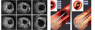
    Because standard IVUS has limitations in distinguishing all plaque components, advanced versions like Virtual Histology IVUS (VH-IVUS) process the ultrasound signals to create a color coded map of the plaque’s composition, showing different types of tissue like fibrous, fatty, and the necrotic core (a dangerous, lipid-rich center within the plaque).


    Optical Coherence Tomography (OCT):

    This is another invasive imaging technique that uses near-infrared light to generate incredibly high resolution images, much finer than what IVUS can provide. OCT is especially good at directly measuring the fibrous cap thickness, which is the thin, protective layer covering the fatty core of the plaque. A fibrous cap that is very thin is a hallmark of a thin cap fibroatheroma, a highly vulnerable plaque.

    OCT can also clearly show lipid rich cores (fatty deposits), signs of inflammation (like macrophage infiltration, where immune cells gather), cholesterol crystals, and small calcium deposits within the plaque, all contributing to a comprehensive assessment of its stability.


    Near Infrared Spectroscopy:

    Often used alongside IVUS, this invasive technique is specifically designed to measure the lipid content within plaques. Near Infrared Spectroscopy creates a chemogram which is essentially a chemical map that highlights areas rich in lipids.


    Other Similar Questions

    Can blood tests help identify vulnerable plaques?

    Yes, certain blood tests can detect biomarkers (biological indicators) related to inflammation and plaque instability, such as C reactive protein (CRP) or specific enzymes called matrix metalloproteinases (MMPs). While useful for general cardiovascular risk assessment, their ability to pinpoint a specific rupture prone plaque is still being researched.


    Is it always necessary to find a vulnerable plaque to assess my heart risk?

    No, doctors also consider your overall cardiovascular risk factors, including age, smoking status, blood pressure, and cholesterol levels, as well as the total amount of plaque throughout your arteries. This comprehensive approach helps assess your vulnerable patient status, which is broader than just finding a single vulnerable plaque.


    If a vulnerable plaque is found, does it always need immediate treatment?

    Not necessarily. The presence of a vulnerable plaque doesn’t automatically mean it will rupture soon. Doctors will evaluate your individual health profile, other risk factors, and the specific characteristics of the plaque to determine the best course of action, which often involves lifestyle changes and medications aimed at stabilizing the plaque.


    Area of Research:

    • Further investigation is focused on the concept of the “vulnerable patient,” which considers overall disease activity and systemic factors in addition to specific plaque features. This aims to improve personalized treatment strategies and potentially target therapies more effectively.

    Resources:

    • Dawson, L. P., & Layland, J. (2022). High-Risk Coronary Plaque Features: A Narrative Review. Cardiology and Therapy, 11(3), 319–335.
    • Gaba, P., Gersh, B. J., Muller, J., Narula, J., & Stone, G. W. (2022). Evolving concepts of the vulnerable atherosclerotic plaque and the vulnerable patient: implications for patient care and future research. Nature Reviews Cardiology.
    • Hafiane, A. (2019). Vulnerable Plaque, Characteristics, Detection, and Potential Therapies. Journal of Cardiovascular Development and Disease, 6(3), 26.
    • Kurihara, O., Takano, M., Miyauchi, Y., Mizuno, K., & Shimizu, W. (2021). Vulnerable atherosclerotic plaque features: findings from coronary imaging. Journal of Geriatric Cardiology, 18(7), 577–584.
    • Sakamoto, A., Cornelissen, A., Sato, Y., Mori, M., Kawakami, R., Kawai, K., Ghosh, S. K. B., Xu, W., Abebe, B. G., Dikongue, A., Kolodgie, F. D., Virmani, R., & Finn, A. V. (2022). Vulnerable Plaque in Patients with Acute Coronary Syndrome: Identification, Importance, and Management. US Cardiology Review, 16, e01.
    • Toutouzas, K., Benetos, G., Karanasos, A., Chatzizisis, Y. S., Giannopoulos, A. A., & Tousoulis, D. (2015). Vulnerable plaque imaging: updates on new pathobiological mechanisms. European Heart Journal, 36(47), 3326–3335.
    • van Veelen, A., van der Sangen, N. M. R., Henriques, J. P. S., & Claessen, B. E. P. M. (2022). Identification and treatment of the vulnerable coronary plaque. Rev. Cardiovasc. Med., 23(1), 039.

  • atherosclerosis image

    What makes an atherosclerotic plaque more vulnerable?

    Overview

    Atherosclerotic plaques are deposits of fatty substances, cholesterol, and other materials that build up inside your arteries. Some atherosclerotic plaques are dangerous because they are more likely to rupture, or break open. When a plaque ruptures, it can lead to the formation of a blood clot (thrombosis) that blocks blood flow, causing serious events like a heart attack (myocardial infarction, MI) or a stroke.

    These plaques are often referred to as vulnerable or high risk plaques because they significantly increase the chance of such life threatening cardiovascular events.

    Understanding what makes a plaque vulnerable is crucial for prevention and treatment. Recent advancements in imaging technologies and ongoing research continue to shed light on the specific characteristics that contribute to a plaque becoming unstable and prone to rupture, helping medical professionals to better assess and manage risk in patients.


    Quick List of Vulnerable Plaque Features

    • Thin Fibrous Cap
    • Large Lipid Core
    • Inflammation and Macrophage Infiltration
    • Spotty or Microcalcifications
    • Intra-plaque Hemorrhage
    • Low or Disturbed Blood Flow
    • Overall Plaque Burden or Volume
    • Positive Remodeling and Napkin Ring Sign

    Thin Fibrous Cap:

    Imagine the plaque as a balloon filled with soft material inside your artery. The fibrous cap is the outer wall of this balloon, acting as a protective layer that separates the plaque’s contents from your bloodstream. A thin fibrous cap is one of the most critical indicators of a vulnerable plaque because if this cap is too thin, it is mechanically weaker and more likely to break or tear. Pathological studies have suggested that plaques with a fibrous cap thickness of less than 65 micrometres (µm) are at high risk of rupture. When this thin cap ruptures, it exposes the highly thrombotic material within the plaque, leading to clot formation.


    Large Lipid Core:

    Inside the fibrous cap, particularly if it’s thin, lies the lipid core (also known as the necrotic core). This core is a collection of fatty material, cholesterol, and dead cells. The larger this lipid-rich core is, the more vulnerable the plaque becomes. This is because the lipid core contains a high amount of tissue factor, a substance that strongly triggers blood clotting. If a large lipid core is exposed to the bloodstream after the fibrous cap ruptures, it dramatically increases the chance of a significant and dangerous blood clot forming.


    Inflammation and Macrophage Infiltration:

    Inflammation plays a central role in the progression and instability of atherosclerotic plaques. Immune cells called macrophages are recruited to the plaque, where they take up lipids and transform into foam cells, contributing to the inflammatory process. These inflammatory cells, particularly macrophages, can produce enzymes (like matrix metalloproteinases) that degrade the fibrous cap, making it thinner and weaker. The presence of macrophages within the fibrous cap is therefore a key feature indicating increased plaque vulnerability and a higher risk of rupture.


    Micro calcification:

    The role of calcium in plaques can be complex. While large, dense areas of calcium are often associated with a more stable, hardened plaque, micro calcifications (very small calcium deposits) within the fibrous cap can actually increase plaque vulnerability. These tiny calcium specks can act as stress concentrators, meaning they create points of weakness in the cap, making it more prone to rupture under mechanical stress. These are early signs of atherosclerosis and can indicate active inflammation. Imaging techniques like coronary CT angiography (CCTA) can detect spotty calcifications, which are often predictive of acute coronary syndrome (ACS).


    Intra Plaque Hemorrhage (IPH):

    Intra plaque Hemorrhage refers to bleeding that occurs within the plaque itself. This internal bleeding can lead to a sudden increase in plaque volume and contributes significantly to the expansion of the lipid core. The accumulation of cholesterol rich red blood cell membranes from the Hemorrhage fuels the growth of the core and further destabilizes the plaque, making it more likely to rupture.


    Flow Dynamics:

    The way blood flows through your arteries exerts force on the vessel walls. Abnormalities in this force can contribute to plaque vulnerability. For instance, when blood flow is slow or turbulent, existing plaques become more prone to rupture or erosion. It can also promote inflammation and intra plaque bleeding. Disturbed blood flow, especially near artery branches, is associated with chronic endothelial activation and can lead to plaque erosion, a type of plaque instability where the surface lining of the artery peels away, exposing underlying tissue and triggering clot formation without a fibrous cap rupture.


    Overall Plaque Burden or Volume:

    While specific features make a plaque vulnerable, the total plaque volume (how much plaque has built up) or plaque burden (the amount of artery wall affected by plaque) is also a strong indicator of overall disease activity and risk. Studies show that plaques leading to acute coronary syndrome (ACS) often have significantly higher total plaque volumes. A larger plaque burden overall is associated with an increased risk of future adverse cardiovascular events.


    Positive Remodeling and Napkin Ring Sign:

    Positive remodeling occurs when the artery expands outwards to accommodate the growing plaque, preventing immediate narrowing of the vessel. While this initially preserves blood flow, it can mask the severity of the plaque within, making it harder to detect visually. The napkin ring sign, observed on CCTA imaging, is a specific pattern indicating a large lipid core surrounded by a high enhancing rim. This sign is strongly associated with thin cap and an increased risk of future adverse cardiovascular events.


    Other Similar Questions:

    • Can diet and exercise help make plaques less vulnerable? Yes, lifestyle changes like an improved diet, regular exercise, and stopping smoking, alongside medications, are crucial in helping to stabilise plaques and reduce their vulnerability.
    • How do doctors identify these vulnerable plaques in living patients? Doctors use advanced imaging techniques such as Optical Coherence Tomography, Coronary Computed Tomography Angiography, Intravascular Ultrasound, and Near Infrared Spectroscopy to look inside the arteries and assess plaque characteristics like cap thickness, lipid content, and inflammation.

    Resources:

    • Hafiane, A. (2019). Vulnerable Plaque, Characteristics, Detection, and Potential Therapies. Journal of Cardiovascular Development and Disease, 6(3), 26.
    • Sakamoto, A., Cornelissen, A., Sato, Y., Mori, M., Kawakami, R., Kawai, K., Ghosh, S. K. B., Xu, W., Abebe, B. G., Dikongue, A., Kolodgie, F. D., Virmani, R., & Finn, A. V. (2022). Vulnerable Plaque in Patients with Acute Coronary Syndrome: Identification, Importance, and Management. US Cardiology Review, 16, e01.
    • Dawson, L. P., & Layland, J. (2022). High-Risk Coronary Plaque Features: A Narrative Review. Cardiology and Therapy, 11(3), 319–335.
    • Kurihara, O., Takano, M., Miyauchi, Y., Mizuno, K., & Shimizu, W. (2021). Vulnerable atherosclerotic plaque features: findings from coronary imaging. Journal of Geriatric Cardiology, 18(7), 577–584.

  • Endothelial Dysfunction image

    What Exactly Triggers Endothelial Dysfunction ?

    Overview

    Your blood vessels are lined by a crucial, dynamic layer of cells called the endothelium. This lining is like a smooth, protective shield that helps keep your blood flowing properly and your arteries healthy in a process known as vascular homeostasis. However, when this lining gets damaged or doesn’t work as it should, it’s called endothelial cell dysfunction (ECD). ECD is a significant contributor to serious conditions like atherosclerosis, which is the hardening and narrowing of arteries, and its complications such as heart attacks and strokes.

    Understanding what triggers endothelial cell dysfunction is important because it’s the earliest detectable change in the development of atherosclerosis and can even predict future heart-related problems. Many factors, from your lifestyle to environmental exposures and underlying health conditions, can lead to endothelial dysfunction, essentially tipping the delicate balance of your blood vessel health towards disease.


    In Details ; What Exactly Triggers Endothelial Dysfunction ?

    One of the earliest and most significant signs of this damage is when endothelial cells start to display adhesion molecules on their surface.

    Think of these adhesion molecules as tiny sticky tags that pop up on the surface of your blood vessel lining. Their appearance is a direct signal of underlying damage or stress to the endothelium, effectively changing the smooth, non stick surface into one that attracts circulating cells and particles. This stickiness is a key first step in the development of serious conditions like atherosclerosis, because it allows inflammatory cells and harmful cholesterol particles to attach and begin building up plaques.


    A quick list of what makes Endothelial cells show their adhesion molecules

    • Pro-inflammatory Cytokines
    • Oxidized Lipoproteins (e.g., Oxidized LDL)
    • Advanced Glycation End Products (AGEs)
    • Disturbed Blood Flow (Hemodynamic Forces)
    • Bacterial Products (e.g., Endotoxins)
    • Certain Viruses
    • Chronic Inflammation
    • Oxidative Stress

    Inflammation and inflammatory cytokines :

    One of the primary triggers for endothelial cells to display adhesion molecules is inflammation. When your body experiences an injury, infection, or chronic stress, certain signaling molecules called pro-inflammatory cytokines (such as Interleukin-1 [IL-1], Tumor Necrosis Factor [TNF], and Interferon-gamma) are released.

    These cytokines act directly on endothelial cells, causing them to become activated. This activation leads to a coordinated program of gene changes within the endothelial cells, turning on the production and display of endothelial-leukocyte adhesion molecules like Vascular Cell Adhesion Molecule-1 (VCAM-1) and Endothelial-Leukocyte Adhesion Molecule-1 (E-selectin), as well as Intercellular Cell Adhesion Molecule-1 (ICAM-1). These molecules act like sticky anchors on the vessel surface, marking it as a site of potential trouble and initiating the recruitment of immune cells.


    Lipoproteins and Advanced Glycation End Products :

    Another major set of triggers comes from the chemical environment of your blood. High cholesterol, particularly elevated levels of bad Low Density Lipoprotein (LDL) cholesterol, plays a significant role. When LDL cholesterol becomes chemically altered, or oxidized , it becomes highly damaging to endothelial cells.

    These oxidized lipoproteins directly stimulate the endothelium to express adhesion molecules like VCAM-1, initiating the process of plaque formation.

    Similarly, in conditions like diabetes with consistently high blood sugar, harmful compounds called Advanced Glycation End Products (AGEs) accumulate in the vessel walls. These AGEs also trigger endothelial cells to switch on the production of adhesion molecules, contributing to the stickiness and furthering the damage.


    Disturbed Blood Flow (Hemodynamic Forces):

    The physical forces of blood flowing through your arteries, known as hemodynamic forces , are also critical triggers. Atherosclerotic plaques don’t form randomly; they tend to appear in specific areas, such as arterial branch points and curves.

    In these regions, blood flow can be disturbed, meaning it’s turbulent or oscillatory, rather than smooth and laminar (undisturbed). This mechanical stress directly causes the endothelial cells to increase the expression of inflammatory adhesion molecules like P-selectin, VCAM-1, and ICAM-1, making these areas more susceptible to immune cell attachment and plaque development.


    The Central Role of NFκB :

    Many of these diverse triggers—whether they are pro-inflammatory cytokines, oxidized lipoproteins, AGEs, or disturbed blood flow—converge on a common internal master switch molecule within the endothelial cells called Nuclear factor-kappa-B (NFκB). NFκB is a pleiotropic transcription factor, meaning it controls the expression of many different genes. When activated by these harmful stimuli, NFκB moves into the cell’s nucleus and instructs the cell to produce more of these adhesion molecules (like VCAM-1 and MCP-1), as well as other pro inflammatory and pro thrombotic substances.

    This makes NFκB a central orchestrator of the endothelial cell’s shift to a damaged, pro-atherosclerotic state, prominently signalled by the increased display of adhesion molecules.


    The Consequence :

    Once these adhesion molecules are expressed on the endothelial surface, they act as specific binding sites for circulating immune cells, primarily monocytes. These immune cells adhere to the sticky endothelial lining and then migrate into the vessel wall (the sub-endothelial space).

    Monocytes can transform into macrophages, which then engulf modified lipoproteins (LDL) and become foam cells – a hallmark of early atherosclerotic lesions. This entire process highlights how the initial appearance of adhesion molecules is a critical, early event in the complex and progressive development of atherosclerosis.


    Other Similar Questions

    Can doctors measure these adhesion molecules to check for vessel damage?

    Yes, increased levels of soluble forms of these adhesion molecules, such as sVCAM-1 and sE-selectin, can be detected in the circulating blood and may serve as markers of endothelial damage and disease severity.


    Resources

    • Gimbrone, M. A., Jr., & García-Cardeña, G. (2016). Endothelial Cell Dysfunction and the Pathobiology of Atherosclerosis. Circulation Research, 118(4), 620–636. doi:10.1161/CIRCRESAHA.115.306301
    • Poredos, P., Poredos, A. V., & Gregoric, I. (2021). Endothelial Dysfunction and Its Clinical Implications. Angiology, 72(7), 604–615. doi:10.1177/0003319720987752
    • Mannarino, E., & Pirro, M. (2008). Endothelial Injury and Repair: A Novel Theory for Atherosclerosis. Angiology, 59(Suppl 2), 69S–72S. doi:10.1177/0003319708320761
    • Wang, X., & He, B. (2024). Endothelial dysfunction: molecular mechanisms and clinical implications. MedComm, 5(e651). doi:10.1002/mco2.651
    • Shimokawa, H. (1999). Primary Endothelial Dysfunction: Atherosclerosis. Journal of Molecular and Cellular Cardiology, 31(1), 23–37. Article No. jmcc.1998.0841
  • Coronary Arteries

    Coronary Arteries

    Overview 

    Your coronary arteries are crucial blood vessels that deliver oxygen-rich blood directly to your heart muscle. Think of them as the heart’s own private plumbing system, ensuring it gets the continuous supply it needs to beat and pump blood throughout your entire body.

    When these arteries become narrowed or blocked, often due to a condition called Coronary Artery Disease (CAD), it can lead to serious problems like a heart attack. Knowing about these arteries and adopting heart healthy habits are key steps in protecting your heart and overall well being.


    In Details

    Here’s a quick list of what we’ll cover:

    • What are coronary arteries?
    • Their function and purpose
    • Normal anatomy and variations
    • Conditions and disorders affecting them
    • Questions & Answers about coronary arteries

    What are coronary arteries? 

    Your coronary arteries are major blood vessels that branch off your aorta, which is the main artery in your body. They are specifically responsible for supplying blood to your heart muscle itself.

    These arteries are located both around and inside your heart muscle, also known as the myocardium. They start at the aortic root, which is the first part of your aorta connected to the left side of your heart.

    The main coronary arteries are usually quite small, typically between 3 and 4 millimetres in diameter. Their exact size can vary slightly depending on factors like your sex, body weight, and ethnicity.


    What is their function and purpose?

    The main job of your coronary arteries is to supply your heart with oxygen rich blood. Your heart is a powerful muscle that needs a constant flow of this blood to work properly and pump blood to the rest of your body. You have two main coronary arteries:

    The right coronary artery (RCA)

    This artery supplies blood to the right upper chamber (right atrium) and the right lower chamber (right ventricle) of your heart. The RCA’s branches also typically supply the sinoatrial (SA) node) and atrioventricular (AV) node), which are like the heart’s natural pacemakers, sending electrical signals that tell your heart muscles when to contract. It also gives blood to one-third of your interventricular septum, which is the wall separating the two lower chambers of your heart. In most people (about 9 out of 10), the RCA is dominant, meaning it supplies a large portion of the heart.

    The left main coronary artery (LMCA)

    This artery supplies blood to the left upper chamber (left atrium) and the left lower chamber (left ventricle). The LMCA’s branches supply the remaining two thirds of your interventricular septum. This artery is usually larger than the right coronary artery and supplies most of the left ventricle and the muscular wall that divides the heart’s lower chambers. The LMCA typically divides into two major branches: the circumflex artery and the anterior interventricular artery (also know as the left anterior descending artery). These two, along with the right coronary artery, are often referred to when discussing “3 vessel coronary arterial disease”.


    Normal anatomy and its variations?

    While there’s a typical arrangement for coronary arteries, their exact layout can vary significantly from person to person. These differences are known as variations or anomalies. For example:

    • Coronary arteries are usually surrounded by a layer of fat, but in some people, they can run directly within the heart muscle itself.
    • The blood supply to the SA and AV nodes usually comes from the right coronary artery , but in about 1 in 10 people (10%), it comes from the left coronary artery instead.
    • Occasionally, only a single coronary artery might come off the aorta, which then divides into the right and left branches.
    • Anatomic variations in the origin and path of coronary arteries are rare, occurring in about 0.3% to 1.6% of individuals based on studies.

    Most of these variations are not harmful and don’t cause any symptoms. However, in very rare cases (less than 1% of people), certain coronary artery abnormalities can lead to serious health problems or even be life threatening. For instance, an interarterial course, where an artery passes between the aorta and pulmonary trunk, is of significant clinical concern as it can be associated with sudden cardiac death.


    What conditions and disorders affect coronary arteries?

    The most common condition affecting your coronary arteries is Coronary Artery Disease (CAD). This often happens because of atherosclerosis, which is a buildup of fatty plaques inside your arteries. When arteries become clogged, they prevent enough blood from reaching your heart, which can lead to a heart attack.

    Another related condition is acute coronary syndrome, a term for situations where there is a sudden and severe reduction in blood flow to the heart through the coronary arteries.

    Less common conditions include:

    • Aneurysms: These are bulges in a blood vessel wall.
    • Anomalous coronary artery: This refers to abnormalities present at birth.
    • Coronary artery spasms: This is a sudden tightening of the muscles in your arteries
    • .Coronary arteriovenous fistulas: Abnormal connections between coronary arteries and veins or heart chambers

    Other similar questions

    • Can babies be born with coronary artery problems? Yes, some people can be born with anomalous coronary arteries, which are abnormalities present at birth, or other congenital heart malformations.
    • How are problems with coronary arteries usually discovered? Many times, coronary artery disease is discovered after someone has a heart attack or undergoes a heart procedure like an angioplasty. Symptoms like chest pain can also indicate a problem.
    • Are all variations in coronary artery anatomy dangerous? No, most variations are not harmful and do not cause symptoms. However, some rare variations, such as an artery taking an “interarterial course” (passing between major arteries), can be clinically significant and are associated with risks like sudden cardiac death.

    Resources

    • Loukas, M., Groat, C., Khangura, R., Owens, D. G., & Anderson, R. H. (2009). The Normal and Abnormal Anatomy of the Coronary Arteries. Clinical Anatomy, 22(1), 114–128.
    • Cleveland Clinic. (n.d.). Coronary Arteries: Function & Anatomy. Cleveland Clinic. (Last reviewed on 2 June 2025)

  • Atherosclerosis image

    What is Atherosclerosis

    Overview

    Atherosclerosis is a chronic, slowly progressing disease where fatty and/or fibrous material builds up in the inner layer of your arteries, called the intima. This build-up forms what are known as atherosclerotic plaques or atheromas, a term derived from a Greek word, reflecting the appearance of the lipid material at the core of these plaques. Over time, these plaques can become more fibrous and accumulate calcium.

    This condition can have serious consequences by impeding blood flow, leading to ischemia (insufficient blood supply to tissues). Plaques can also break open, triggering the formation of a thrombus (blood clot) that can block the artery, causing acute ischemia.

    Coronary artery disease (CVD) is a leading cause of vascular disease and death worldwide, significantly contributing to conditions like heart attacks and strokes. Despite advancements in medical care, it remains a major global health challenge.


    In Details

    First: Key Characteristics of Atherosclerosis

    • Build up of fatty and fibrous material: Occurs in the inner lining of arteries.
    • Formation of plaques (atheromas): These are the distinctive lesions of the disease.
    • Narrowing of arteries: Advanced plaques can reduce the space for blood flow.
    • Risk of blood clot formation: Plaques can rupture or erode, leading to clots that block blood flow.
    • Involves Low-Density Lipoprotein (LDL): A key factor in plaque development.
    • Inflammation and other risk factors: Play significant roles in its development.

    Initiation of Atherosclerosis

    Atherosclerosis often begins as early as childhood, though symptoms may not appear for decades. The process is thought to start with damage or injury to the inner layer of an artery, called the endothelium. This damage can be caused by various factors, including high blood pressure, high cholesterol, high triglycerides (another type of fat in the blood), smoking or other tobacco use, diabetes, insulin resistance, obesity, and inflammation. When the artery wall is damaged, low-density lipoprotein (LDL) particles – which transport cholesterol through the blood – can accumulate in the intima, the innermost layer of the artery

    Research strongly suggests that atherosclerosis would likely not occur if LDL-C (low-density lipoprotein cholesterol) concentrations did not exceed the body’s physiological needs. Prolonged exposure to high LDL-C levels over many years is a primary driver of the disease’s initiation and progression. For instance, individuals with familial hypercholesterolaemia (a genetic condition causing very high LDL-C from an early age) develop premature atherosclerotic CVD, highlighting LDL’s causal role.

    Progression of Atherosclerosis

    In its early stages, atherosclerotic plaques often expand outwards, away from the arterial lumen (the hollow centre where blood flows), which helps preserve the artery’s width. However, eventually, the growing plaque begins to encroach upon the arterial lumen, leading to stenosis (narrowing) and potentially flow-limiting lesions. This reduced blood flow can cause symptoms like angina pectoris (chest pain from insufficient blood to the heart) during physical exertion.

    Complications of Atherosclerosis

    The most severe complications of atherosclerosis often arise from the disruption of plaques. The most common trigger for acute coronary syndromes, such as a myocardial infarction (heart attack), is the rupture of an atherosclerotic plaque.

    These “vulnerable plaques” typically have a large lipid core covered by a thin fibrous cap (a layer of fibrous tissue). When this cap breaks, the plaque’s thrombogenic (clot-forming) contents are exposed to the blood, rapidly triggering thrombosis (blood clot formation), which can completely block the artery.

    Another mechanism, increasingly recognized, is plaque erosion, where lesions without a thin cap or much lipid can also lead to clot formation, particularly in the current era of effective anti-atherosclerotic therapies. These clots can lead to acute ischemic events like heart attacks or strokes.


    Other Similar Questions

    What are the common symptoms of atherosclerosis?

    Mild atherosclerosis often causes no symptoms for many years. Symptoms usually only appear when an artery is significantly narrowed or blocked, or when a blood clot forms. Symptoms vary depending on which arteries are affected. For example, chest pain (angina) if it’s in heart arteries, sudden weakness or trouble speaking if it’s in brain arteries (like a stroke), or leg pain when walking (claudication) if it’s in leg arteries.

    Is atherosclerosis curable?

    While atherosclerosis is a chronic and progressive disease, its progression can often be prevented, and in some cases, even reversed, through lifestyle changes and medical treatments. Current treatments aim to manage the disease and prevent its serious complications like heart attacks and strokes.

    How is atherosclerosis diagnosed?

    The diagnosis of atherosclerosis often involves imaging tests that can directly visualise the plaques or assess if there is reduced blood flow to organs. Non-invasive tests include ultrasonography (using sound waves) and CT angiography (a type of X-ray scan).

    More invasive procedures like invasive angiography (using a dye to visualize arteries) might be used to guide treatments.


    Resources

    • “Atherosclerosis” by Peter Libby, Julie E. Buring, Lina Badimon, Göran K. Hansson, John Deanfield, Márcio Sommer Bittencourt, Lale Tokgözoğlu and Eldrin F. Lewis.
    • Arteriosclerosis / atherosclerosis
  • Prevention of Coronary Artery Disease image

    Prevention of Coronary Artery Disease

    Overview

    Prevention of Coronary Artery Disease by modifying what are called modifiable risk factors, which are factors you can change, such as diet, exercise, and smoking habits. Prevention is divided into three key categories: primary, secondary, and tertiary. These approaches aim to stop Coronary Artery Disease before it starts, slow its progression, or manage its impact to improve your quality of life.


    In Details

    • Primary Prevention: Aims to prevent Coronary Artery Disease before it even occurs, for people with risk factors but no symptoms.
    • Secondary Prevention: Focuses on people who already have established Coronary Artery Disease, to prevent further progression and reduce the impact of the disease.
    • Tertiary Prevention: Applies to people with existing Coronary Artery Disease, aiming to improve their quality of life by reducing disability, delaying complications, and restoring heart function.

    Risk factors for Coronary Artery Disease are broadly divided into non-modifiable and modifiable. Non-modifiable factors, which cannot be changed, include age, gender, race, and your genes.

    Modifiable risk factors, which you can influence, include dyslipidemia (unhealthy levels of fats in your blood), diabetes, hypertension, cigarette smoking, obesity, chronic kidney disease, chronic infection, high C-reactive protein (CRP) (a marker of inflammation), hyperhomocysteinemia (HHcy) (high levels of an amino acid called homocysteine), advanced glycation end products (AGEs), oxidative stress, and caffeine. Modifying these factors can prevent, reverse, or slow the progression of Coronary Artery Disease and improve your quality of life.


    Primary Prevention of Coronary Artery Disease 

    Primary prevention is about taking steps to prevent the start of atherosclerosis. This involves assessing your risk, especially if you are between 40 and 75 years of age.

    Coronary Artery Disease risk score, like the one developed by the American College of Cardiology ASCVD risk estimator, considers factors such as age, sex, race, cholesterol levels, blood pressure, diabetes, smoking, and medication use to estimate your 10-year risk of having Coronary Artery Disease or a stroke. This score, expressed as a percentage, helps determine if you are at low (0.0-4.9%), borderline (5.0-7.4%), intermediate (7.5-20.0%), or high risk (>20.0%) and guides prevention strategies.

    Key interventions for primary prevention include

    • Adopting a Healthy Diet: This means consuming plenty of fruits, vegetables, nuts, whole grains, lean proteins (vegetable or animal), and fish.
      It’s advised to reduce intake of trans fats, red meat, processed meat, added sugars, saturated fat, sweetened beverages, and sodium. Foods like flaxseed, whey protein, and grapefruit (containing resveratrol) can help lower “bad cholesterol” and raise “good cholesterol”.
    • Managing AGE-RAGE Stress: AGEs are harmful compounds that can build up in the body and contribute to atherosclerosis. They are found in foods like red meat, cheese, cream, animal fat, and sweetened pastries, so reducing their consumption is advised.
    • Reducing Oxidative Stress: This occurs when there’s an imbalance between harmful reactive oxygen species (ROS) (unstable molecules that can damage cells) and your body’s antioxidants.
      Eating antioxidant-rich foods like flaxseed, fruits, and vegetables can help. Limiting heavy alcohol consumption is also recommended.
    • Regular Physical Activity: Aim for 150 minutes per week of moderate-intensity physical activity or 75 minutes per week of high-intensity physical activity.
    • Stopping Tobacco Use: Quitting smoking is crucial.
    • Controlling Diabetes and Hypertension: If you have diabetes, managing your diet, exercising, and taking medication as needed are important.
      For hypertension, non-pharmacological interventions are advised, and pharmacological therapy may be recommended to achieve a blood pressure target of less than 130/80 mmHg.
    • Aspirin Use: Infrequent use of aspirin may be advised.

    Secondary Prevention of Coronary Artery Disease

     Secondary prevention is for individuals who have already been diagnosed with Coronary Artery Disease, have current symptoms, or have experienced a heart attack, or undergone procedures like percutaneous coronary intervention (PCI) (a procedure to open blocked arteries, often using a balloon and stent) or coronary artery bypass graft (CABG) (a surgical procedure to create new pathways for blood flow around blocked arteries).
    The main goal is to slow or reverse the disease’s progression and reduce the risk of future events or death.

    Key interventions for secondary prevention include

    • Lifestyle Modification: This includes diet similar to primary prevention. Physical activity recommendations are similar, with a target of at least 150 minutes per week of moderate-intensity or 75 minutes per week of high-intensity physical activity. Stopping cigarette smoking is also critical.
    • Serum Lipids Management: Medications like statins are used to lower LDL-C. Fibrates or high doses of omega-3 fatty acids might be used if triglyceride (TG) levels are high.
    • Hypertension Management: Antihypertensive drugs may be used. Sodium and alcohol restriction are also important, with a blood pressure target of 130/80 mmHg.
    • Antiplatelet Drugs: If your 10-year risk score is 10% or higher, antiplatelet drugs like aspirin or clopidogrel may be used to prevent blood clotting.
    • Diabetes Management: The aim is to normalise blood glucose levels using antidiabetic drugs, with a target for HbA1c (a measure of average blood sugar over 2-3 months) of less than 7%.

    Tertiary Prevention of Coronary Artery Disease

     Tertiary prevention is for people with established Coronary Artery Disease, focusing on improving their quality of life by reducing disability, delaying or limiting complications, and restoring heart function.

    This involves a team approach with healthcare professionals like cardiologists, cardiac surgeons, exercise specialists, physiotherapists, dietitians, and nurses. Treatment often includes medical procedures such as PCI, CABG, pacemaker implantation, defibrillator placement, and ventricular-assisted devices.


    Other Similar Questions

    Is Coronary Artery Disease preventable in all cases?

    While it’s largely preventable through risk factor modification, some non-modifiable risk factors like age and genetics mean it might not be entirely avoidable for everyone, but its progression can be significantly slowed.


    Resources

  • Symptoms of Coronary Artery Disease image

    Symptoms of Coronary Artery Disease

    Overview

    Understanding these symptoms is crucial because early detection and management can significantly improve outcomes. The way Coronary Artery Disease presents can vary greatly among individuals, ranging from no noticeable symptoms to severe and life threatening events. It’s important to be aware of the different ways this condition can manifest, as recognizing them promptly can lead to timely medical attention.


    Symptoms of Coronary Artery Disease

    Here is a quick list of common symptoms associated with Coronary Artery Disease:

    • Chest pain or discomfort (Angina Pectoris)
    • Shortness of breath (Dyspnea)
    • Fatigue
    • Nausea
    • Sweating (Diaphoresis)
    • Fainting (Syncope)
    • Palpitations (a feeling of your heart pounding or racing)
    • Cardiac arrest or sudden cardiac death
    • Atypical symptoms, especially in women, the elderly, and diabetics
    • Silent ischemia (no symptoms at all)

    In Details:

    1. Angina pectoris

    The most common symptom of Coronary Artery Disease is angina pectoris, often simply called angina. This is a clinical syndrome characterized by discomfort in the chest. People often describe it as a tight, squeezing, heavy, or pressure like feeling, rather than a sharp pain. This discomfort can be felt in the center of the chest (substernal) and may spread or radiate to other areas, such as the jaw, shoulder, back, arms (especially the left arm, or both arms), or even the teeth and upper abdomen (epigastric region).

    Angina is typically brought on by increased demand on the heart, such as during physical exertion, emotional stress, after a heavy meal, or exposure to cold. It usually lasts for several minutes and can be relieved by rest or by taking medication like sublingual nitroglycerin.


    2. Dyspnea

    Another significant symptom is dyspnea, or shortness of breath. More commonly, shortness of breath occurs alongside chest discomfort, described as a feeling of tightness across the chest or a restriction in breathing. Dyspnea can also indicate more advanced Coronary Artery Disease, such as if there’s ischemic left ventricular dysfunction (when the heart’s main pumping chamber is weakened due to lack of blood flow), or other complications like pulmonary venous congestion or pulmonary oedema.


    3. General symptoms

    Beyond chest discomfort and breathlessness, Coronary Artery Disease can manifest with other general symptoms. These can include nausea, sweating (diaphoresis), and fatigue. Fatigue is particularly common in patients with Coronary Artery Disease, sometimes due to the psychological impact of the disease or when combined with cardiac failure. Some individuals might experience palpitations, a sensation of their heart pounding or racing, or episodes of syncope (fainting).


    4. Atypical symptoms

    It’s important to be aware of atypical symptoms, which are more frequently observed in certain populations like women, the elderly, and individuals with diabetes mellitus. In these groups, Coronary Artery Disease might not present with the classic chest pain. Instead, it could appear as isolated symptoms such as palpitations, extreme fatigue, unusual discomfort (e.g., in the neck, jaw, or back without chest pain), or even lead directly to cardiac arrest without prior noticeable signs. The diagnosis of these atypical presentations can be challenging and requires careful clinical assessment.


    5. Silent ischemia

    A particularly concerning aspect of Coronary Artery Disease is silent ischemia. This refers to episodes where the heart muscle is not receiving enough blood (ischemia) but the person experiences no pain or discomfort at all. This lack of symptoms means that significant heart damage can occur without any warning. Silent ischemia is more prevalent among the elderly and people with diabetes mellitus.

    In some tragic cases, the very first manifestation of Coronary Artery Disease is a heart attack (myocardial infarction), which can be fatal. In fact, for approximately one in four people, the first symptom of coronary artery disease is what is termed sudden cardiac death. In these instances, there may be no prior warning signs or symptoms at all. However, for some who experience cardiac arrest, there might have been “warning symptoms” like chest pain or dyspnea in the days or weeks leading up to the event.


    Other similar questions

    Can I have coronary artery disease without any symptoms?

    Yes, it is possible. Many individuals, especially the elderly and those with diabetes, can have what is called “silent ischemia,” where they experience reduced blood flow to the heart without any noticeable pain or discomfort

    Do men and women experience Coronary Artery Disease symptoms differently?

    While the core symptoms are similar, women, the elderly, and diabetics are more prone to “atypical symptoms” which might not include classic chest pain. These can manifest as fatigue, shortness of breath, nausea, or discomfort in areas like the jaw, neck, or back, rather than the chest.

    When to seek medical attention ?

    If you experience symptoms that suggest Coronary Artery Disease, such as chest pain or discomfort, shortness of breath, or unexplained fatigue, you should seek medical attention promptly. For severe or sudden symptoms like crushing chest pain, especially if it radiates or is accompanied by sweating or nausea, call for emergency medical help immediately.


    Resources

    • Malakar, A. K., Choudhury, D., Halder, B., Paul, P., Uddin, A., & Chakraborty, S. (2019). A review on coronary artery disease, its risk factors, and therapeutics. Journal of Cellular Physiology, 234(10), 16812–16824.
    • Bergmark, B. A., Mathenge, N., Merlini, P. A., Lawrence-Wright, M. B., & Giugliano, R. P. (2021). Acute coronary syndromes. The Lancet, 398(10300), 741–756.
    • Smith, J. N., Negrelli, J. M., Manek, M. B., Hawes, E. M., & Viera, A. J. (2015). Diagnosis and Management of Acute Coronary Syndrome: An Evidence-Based Update. Journal of the American Board of Family Medicine, 28(2), 283–293.
    • Geller, B. J., & Abella, B. S. (2018). Evolving Strategies in Cardiac Arrest Management. Cardiology Clinics, 36(1), 73–84.
    • Granfeldt, A. (2019). In-Hospital Cardiac Arrest. JAMA, 321(16), 1618-1619.
    • Krahn, A. D., Tfelt-Hansen, J., Tadros, R., Steinberg, C., Semsarian, C., & Han, H.-C. (2022). Latent Causes of Sudden Cardiac Arrest. JACC: Clinical Electrophysiology, 8(6), 806–821.
    • Gallone, G., Baldetti, L., Pagnesi, M., Latib, A., Colombo, A., Libby, P., & Giannini, F. (2018). Medical Therapy for Long-Term Prevention of Atherothrombosis Following an Acute Coronary Syndrome. Journal of the American College of Cardiology, 72(23), 2886–2903.
    • Bahit, M. C., Korjian, S., Daaboul, Y., Baron, S., Bhatt, D. L., Kalayci, A., Chi, G., Nara, P., Shaunik, A., & Gibson, C. M. (2023). Patient Adherence to Secondary Prevention Therapies After an Acute Coronary Syndrome: A Scoping Review. Clinical Therapeutics, 45(9), 1119–1126.
    • Gaviria-Mendoza, A., Zapata-Carmona, J. A., Restrepo-Bastidas, A. A., Betancur-Pulgarín, C. L., & Machado-Alba, J. E. (2020). Prior Use of Medication for Primary Prevention in Patients with Coronary Syndrome. Journal of Primary Care & Community Health, 11, 2150132720946949.
    • Silverio, A., Cancro, F. P., Esposito, L., Bellino, M., D’Elia, D., Verdoia, M., Vassallo, M. G., Ciccarelli, M., Vecchione, C., Galasso, G., & De Luca, G. (2023). Secondary Cardiovascular Prevention after Acute Coronary Syndrome: Emerging Risk Factors and Novel Therapeutic Targets. Journal of Clinical Medicine, 12(6), 2161.
    • Fitchett, D. H., Leiter, L. A., Lin, P., Pickering, J., Welsh, R., Stone, J., Gregoire, J., McFarlane, P., Langer, A., Gupta, A., & Goodman, S. G. (2020). Update to Evidence-Based Secondary Prevention Strategies After Acute Coronary Syndrome. CJC Open, 2(4), 402–415.
    • Isted, A., Williams, R., & Oakeshott, P. (2018). Secondary prevention following myocardial infarction: A clinical update. British Journal of General Practice, 68(669), 151–152.
    • Bavishi, A., Howard, T., Ho-Kim, J., Hiramato, B., Pierce, J. B., Mendapara, P., Alhalel, J., Wu, H.-W., Srdanovich, N., & Stone, N. (2018). Treatment Gap in Primary Prevention Patients Presenting with Acute Coronary Syndrome. The American Journal of Cardiology, 123(2), 237–242.
    • Sun, Z. (2013). Cardiac Imaging Modalities in the Diagnosis of Coronary Artery Disease. Journal of Clinical & Experimental Cardiology, S6(e001), 1–4.
    • Ford, T. J., & Berry, C. (2020). Angina: Contemporary diagnosis and management. Heart, 106(5), 387–398.
    • Libby, P., & Theroux, P. (2005). Pathophysiology of Coronary Artery Disease. Circulation, 111(25), 3481–3488.
    • Mayo Clinic. (n.d.). Coronary artery disease – Diagnosis and treatment. Retrieved from.
    • Albus, C., Barkhausen, J., Fleck, E., Haasenritter, J., Lindner, O., & Silber, S. (2017). The Diagnosis of Chronic Coronary Heart Disease. Deutsches Ärzteblatt International, 114(42), 712–719.
  • Complications of Coronary Artery Disease

    Complications of Coronary Artery Disease

    Overview

    Understanding the potential complications of Coronary Artery Disease is crucial for patients and their loved ones, as it highlights the importance of early diagnosis, effective management, and adopting healthy lifestyle.

    Coronary Artery Disease itself is an atherosclerotic disease, meaning it involves the buildup of plaque inside the arteries, which is inflammatory in nature. This plaque accumulation starts when the lining of the arterial wall is disrupted, leading to lipoprotein droplets gathering in the coronary vessels. Over time, these plaques can disrupt or erode, potentially leading to serious complications.


    In Detail
    Complications of Coronary Artery Disease

    Here’s a quick list of the main complications of Coronary Artery Disease:

    The manifestations and complications of Coronary Artery Disease can vary, from less severe symptoms to life threatening events.


    Angina Pectoris

    Angina Pectoris, or chest pain, is a common clinical syndrome associated with myocardial ischemia. This discomfort can be felt in the chest, jaw, arm, or other areas. While often described as a tight, squeezing, or heavy feeling, its localization can be vague, and there’s considerable individual variation. Angina can be stable, unstable, or even manifest as variant angina (Prinzmetal angina) or microvascular angina. It is important to know that many patients experiencing angina, about half, do not have obstructive coronary artery disease (blockages visible on angiography) but rather have microvascular angina (MVA), which involves problems with the very small blood vessels of the heart, and/or vasospastic angina (VSA), caused by sudden narrowing or spasm of the heart arteries. A critical point is that angina symptoms do not always directly correlate with the severity of the underlying atherosclerosis, and some patients can have considerable myocardial ischemia without any pain, a condition known as ‘silent ischemia’, which is more common in the elderly and individuals with diabetes mellitus.


    Myocardial Infarction (MI) or heart attack

    One of the most serious complications is a Myocardial Infarction (MI), commonly known as a heart attack. This occurs when there is evidence of myocardial necrosis (heart muscle death) due to acute myocardial ischemia, meaning the heart muscle doesn’t get enough blood flow. A heart attack can be classified as either an ST-segment elevation myocardial infarction (STEMI), a severe type where there’s a complete blockage of a heart artery, or a non-ST-segment elevation myocardial infarction (NSTEMI), where blood flow is severely reduced but not completely blocked. For about half of people, a heart attack is the very first symptom they experience from Coronary Artery Disease, and sadly, half of these initial heart attacks can be fatal. Heart attacks are associated with substantial morbidity and mortality and can even lead to mechanical complications such as acute ventricular septal rupture (a hole in the heart wall), acute mitral regurgitation (a leaky heart valve), or free wall rupture. Previous research also indicates that recurrent heart attacks are linked to worse outcomes than the initial event.


    Sudden Cardiac Death (SCD)

    Another devastating complication is Sudden Cardiac Death (SCD), also known as Cardiac Arrest (CA). In fact, for one out of four people, sudden cardiac death is the very first symptom of coronary artery disease. Coronary artery disease is the most common cause of cardiac arrest, accounting for over 80% of all cases. The prognosis for sudden cardiac arrest remains poor, with a survival rate to hospital discharge typically between 6% and 10%. Many individuals unfortunately die before even reaching the hospital. Even among those successfully resuscitated, there is a high rate of in-hospital mortality. In survivors of cardiac arrest, a post resuscitation syndrome is commonly observed, which frequently includes transient myocardial dysfunction.


    Arrhythmias

    Coronary artery disease can also lead to Arrhythmias, which are irregular heartbeats. For instance, atrial fibrillation, a common type of irregular heartbeat where the upper chambers of the heart beat rapidly and irregularly, is observed in 20% of patients with Coronary artery disease. More severe arrhythmias, such as ventricular tachycardia (a fast, abnormal heart rhythm starting in the lower chambers of the heart), can also occur. These irregular heartbeats can sometimes lead to complications like atrioventricular block or, in the most severe cases, sudden collapse and death.


    Heart failure (HF)

    Heart failure (HF), a condition where the heart cannot pump enough blood to meet the body’s needs, is another common consequence of Coronary artery disease. This often indicates advanced coronary artery disease, perhaps due to a prior heart attack or widespread myocardial fibrosis (scarring of the heart muscle) resulting from previous ischemic episodes.


    In some cases, Coronary artery disease may present with peripheral arterial embolism (a blockage of an artery in the arms or legs) and embolic stroke (a type of stroke caused by a blood clot traveling to the brain), typically following a heart attack where a blood clot forms within the heart’s left ventricle.

    The pervasive impact of Coronary artery disease extends beyond individual health to society at large. Patients with conditions like microvascular angina and vasospastic angina often experience a profound and long-term impact on their physical and mental well-being, leading to repeated hospitalizations and a reduced quality of life. This underscores the critical need for effective prevention and management strategies.


    Other Similar Questions

    How does Coronary artery disease affect the body beyond the heart?

    Coronary artery disease can lead to issues such as peripheral arterial embolism and embolic stroke, usually after a heart attack. It can also contribute to overall decreased quality of life due to chronic symptoms like fatigue and dyspnea.

    Can Coronary artery disease be present without any symptoms?

    Yes, it is possible to have significant myocardial ischemia without experiencing any symptoms, a condition known as ‘silent ischemia.’ This is more common in elderly patients and those with diabetes mellitus.

    Why is it important for family members to be aware of Coronary artery disease complications?

    Because sudden cardiac death can be the first manifestation of Coronary artery disease, and many cases are unexplained even after investigation, family screening for inherited disorders, especially when sudden death occurs before 40-45 years of age, is important to identify at-risk individuals. Awareness of symptoms and risk factors can prompt earlier medical attention.


    Resources

    • Malakar, A. K., Choudhury, D., Halder, B., Paul, P., Uddin, A., & Chakraborty, S. (2019). A review on coronary artery disease, its risk factors, and therapeutics. Journal of Cellular Physiology, 234(10), 16812–16824.
    • Bergmark, B. A., Mathenge, N., Merlini, P. A., Lawrence-Wright, M. B., & Giugliano, R. P. (2021). Acute coronary syndromes. The Lancet, 398(10300), 741–756.
    • Smith, J. N., Negrelli, J. M., Manek, M. B., Hawes, E. M., & Viera, A. J. (2015). Diagnosis and Management of Acute Coronary Syndrome: An Evidence-Based Update. Journal of the American Board of Family Medicine, 28(2), 283–293.
    • Geller, B. J., & Abella, B. S. (2018). Evolving Strategies in Cardiac Arrest Management. Cardiology Clinics, 36(1), 73–84.
    • Granfeldt, A. (2019). In-Hospital Cardiac Arrest. JAMA, 321(16), 1618-1619.
    • Krahn, A. D., Tfelt-Hansen, J., Tadros, R., Steinberg, C., Semsarian, C., & Han, H.-C. (2022). Latent Causes of Sudden Cardiac Arrest. JACC: Clinical Electrophysiology, 8(6), 806–821.
    • Gallone, G., Baldetti, L., Pagnesi, M., Latib, A., Colombo, A., Libby, P., & Giannini, F. (2018). Medical Therapy for Long-Term Prevention of Atherothrombosis Following an Acute Coronary Syndrome. Journal of the American College of Cardiology, 72(23), 2886–2903.
    • Bahit, M. C., Korjian, S., Daaboul, Y., Baron, S., Bhatt, D. L., Kalayci, A., Chi, G., Nara, P., Shaunik, A., & Gibson, C. M. (2023). Patient Adherence to Secondary Prevention Therapies After an Acute Coronary Syndrome: A Scoping Review. Clinical Therapeutics, 45(9), 1119–1126.
    • Gaviria-Mendoza, A., Zapata-Carmona, J. A., Restrepo-Bastidas, A. A., Betancur-Pulgarín, C. L., & Machado-Alba, J. E. (2020). Prior Use of Medication for Primary Prevention in Patients with Coronary Syndrome. Journal of Primary Care & Community Health, 11, 2150132720946949.
    • Silverio, A., Cancro, F. P., Esposito, L., Bellino, M., D’Elia, D., Verdoia, M., Vassallo, M. G., Ciccarelli, M., Vecchione, C., Galasso, G., & De Luca, G. (2023). Secondary Cardiovascular Prevention after Acute Coronary Syndrome: Emerging Risk Factors and Novel Therapeutic Targets. Journal of Clinical Medicine, 12(6), 2161.
    • Fitchett, D. H., Leiter, L. A., Lin, P., Pickering, J., Welsh, R., Stone, J., Gregoire, J., McFarlane, P., Langer, A., Gupta, A., & Goodman, S. G. (2020). Update to Evidence-Based Secondary Prevention Strategies After Acute Coronary Syndrome. CJC Open, 2(4), 402–415.
    • Isted, A., Williams, R., & Oakeshott, P. (2018). Secondary prevention following myocardial infarction: A clinical update. British Journal of General Practice, 68(669), 151–152.
    • Bavishi, A., Howard, T., Ho-Kim, J., Hiramato, B., Pierce, J. B., Mendapara, P., Alhalel, J., Wu, H.-W., Srdanovich, N., & Stone, N. (2018). Treatment Gap in Primary Prevention Patients Presenting with Acute Coronary Syndrome. The American Journal of Cardiology, 123(2), 237–242.
    • Sun, Z. (2013). Cardiac Imaging Modalities in the Diagnosis of Coronary Artery Disease. Journal of Clinical & Experimental Cardiology, S6(e001), 1–4.
    • Ford, T. J., & Berry, C. (2020). Angina: Contemporary diagnosis and management. Heart, 106(5), 387–398.
    • Libby, P., & Theroux, P. (2005). Pathophysiology of Coronary Artery Disease. Circulation, 111(25), 3481–3488.
    • Mayo Clinic. (n.d.). Coronary artery disease – Diagnosis and treatment. Retrieved from.
    • Albus, C., Barkhausen, J., Fleck, E., Haasenritter, J., Lindner, O., & Silber, S. (2017). The Diagnosis of Chronic Coronary Heart Disease. Deutsches Ärzteblatt International, 114(42), 712–719.
  • What is Cardiac arrest?

    What is Cardiac Arrest?

    Overview

    What is Cardiac Arrest? Cardiac arrest is a sudden and often unexpected event where the heart abruptly stops effectively pumping blood throughout the body. This leads to an immediate loss of circulation, which is why it is so critical.

    Despite extensive research and investment in the field, the outlook for sudden cardiac arrest remains poor, with a survival rate to hospital discharge typically between 6% and 10%. Most people who experience cardiac arrest sadly do not even reach the hospital. Improving outcomes for these patients requires focused efforts at every stage of their care, with heart-related issues being central to this challenge, from before the event occurs, throughout resuscitation, and into the long term follow up for survivors.


    In Details
    What is Cardiac Arrest

    Cardiac arrest is a complex medical emergency. Here’s a quick look at some key aspects:

    • Often caused by pre-existing heart disease.
    • Management involves immediate life support, specialized medical care, and ongoing follow up.
    • Prevention strategies are crucial, both for individuals at high risk and within hospital settings.

    Understanding Cardiac Arrest

    Cardiac arrest is defined as the abrupt and often unanticipated cessation of cardiac output (when the heart stops pumping blood effectively). It is also described as the loss of circulation prompting resuscitation with chest compressions, defibrillation, or both. This means the heart has stopped its vital pumping function, leading to a sudden and complete lack of blood flow to the brain and other organs. If not immediately treated, it can be fatal.


    What Causes Cardiac Arrest?

    Many factors can lead to cardiac arrest:

    • Pre existing heart disease is a common underlying factor.
    • Coronary artery disease (CAD), which affects the blood vessels supplying the heart, is the most common cardiac cause, accounting for over 80% of cases due to heart related issues, especially in men. Acute myocardial ischemia (a lack of blood flow to the heart muscle) is a prominent contributor.
    • Other cardiac causes include structural heart diseases like dilated cardiomyopathy (enlarged heart muscle) or hypertrophic cardiomyopathy (thickened heart muscle), and electrophysiological diseases (conditions affecting the heart’s electrical system) such as Brugada’s syndrome or long QT syndrome. These non-ischemic conditions make up less than 20% of primary cardiac causes.
    • The causes can vary with age; for instance, ischemic cardiomyopathy is more common in patients over 35, while a wider range of causes are seen in those under 35.
    • In hospital cardiac arrest (IHCA), where the event occurs while a patient is in hospital, cardiac causes are still the most frequent (50-60%), followed by respiratory insufficiency (15-40%).
    • Sometimes, despite thorough medical investigation, the cause remains unexplained, partly due to the high mortality before reaching hospital and low autopsy rates.
    • Latent causes (hidden or less obvious causes) are also being increasingly recognised. These include inherited arrhythmia syndromes such as catecholaminergic polymorphic ventricular tachycardia (CPVT), short QT syndrome (SQTS), early repolarization syndrome (ERS), and short-coupled ventricular fibrillation (SCVF). These can account for a significant portion of previously “unexplained” cardiac arrests.

    Can Cardiac Arrest Be Prevented?

    Yes, efforts are made to prevent cardiac arrest:

    • Primary prevention aims to identify high-risk individuals (those with known heart disease) before an event occurs. For some, an implantable cardiac defibrillator (ICD), a small device surgically placed in the body to monitor heart rhythm and deliver an electrical shock if needed, can be used. However, effective tools for identifying at-risk individuals in the general population are still needed.
    • Subacute prevention involves recognizing “warning symptoms,” such as chest pain or shortness of breath, which may appear days or weeks before a cardiac arrest. This suggests that “sudden death is not so sudden” in all cases, potentially allowing for early intervention.
    • In hospitals, prevention includes systems to identify deteriorating patients and the use of rapid response teams. Addressing medication-related issues and managing conditions like sepsis can also help prevent IHCA.
    • Familial screening is also important for first-degree relatives of sudden death victims, especially when the event occurs before age 40-45, to identify inherited heart conditions.

    How is Cardiac Arrest Managed?

    Management of cardiac arrest is divided into “during” and “after” the event:

    • During Cardiac Arrest
      • The core of immediate treatment involves chest compressions, ventilation (providing breaths), and early defibrillation (an electrical shock to reset the heart’s rhythm) if the heart rhythm is “shockable”.
    • For cardiac arrest caused by a heart attack (acute myocardial infarction), an early invasive strategy, such as immediate coronary angiography (a procedure to visualise the heart’s blood vessels) and percutaneous coronary intervention (PCI) (a procedure to open blocked arteries), is often recommended.
    • High-quality and uninterrupted chest compressions are crucial. For laypersons, focusing on compression-only CPR (Cardiopulmonary Resuscitation) is often advised.
    • Medications like epinephrine are commonly used, though their overall long-term benefit is debated. Amiodarone or lidocaine may be used for shock-resistant heart rhythms.
    • Monitoring the quality of CPR using measures like end-tidal carbon dioxide (ETCO2) levels (a measure of carbon dioxide in exhaled breath, reflecting blood flow) is important, with a goal of at least 20 mmHg.
    • For very severe cases, Extracorporeal Membrane Oxygenation (ECMO), a life support system that takes over the function of the heart and lungs, may be considered, especially if a reversible cause for the arrest can be identified.

    • After Cardiac Arrest (Post-resuscitation Care)
      • Once the heart restarts (Return of Spontaneous Circulation ), care focuses on treating the underlying cause, supporting vital organs, and protecting the brain.
      • Postresuscitation myocardial dysfunction (PRMD), a severe but temporary weakening of the heart muscle, is common and usually resolves within 48 to 72 hours. It is managed with medications to support heart function (like dobutamine).
      • For survivors, secondary prevention is vital to prevent future cardiac events. This includes optimizing treatment for existing heart conditions, using medications like beta-blockers, and often implanting an implantable cardiac defibrillator to prevent recurrence of life-threatening arrhythmias.
      • Targeted Temperature Management (TTM), which involves carefully controlling the patient’s body temperature, is a primary strategy to protect the brain, typically maintained between 32°C and 36°C for at least 24 hours.

    Other Similar Questions

    Cardiac arrest vs heart attack?

    A heart attack (acute myocardial infarction) is a common cause of cardiac arrest, but they are not the same event. A heart attack occurs when blood flow to a part of the heart muscle is blocked, while cardiac arrest is when the heart stops pumping blood effectively.

    Can cardiac arrest be prevented?

    Yes, in many cases, by identifying and managing pre-existing heart conditions, recognizing early warning signs, and implementing quick response systems, particularly in hospital settings.


    Resources

    Bougouin, W., & Cariou, A. (2017). Cardiac Issues in Cardiac Arrest. Seminars in Neurology, 37(01), 13–18. Geller, B. J., & Abella, B. S. (2018). Evolving Strategies in Cardiac Arrest Management. Cardiology Clinics, 36(1), 73–84. Andersen, L. W., Holmberg, M. J., Berg, K. M., Donnino, M. W., & Granfeldt, A. (2019). In-Hospital Cardiac Arrest: A Review. JAMA, 321(12), 1200. Krahn, A. D., Tfelt-Hansen, J., Tadros, R., Steinberg, C., Semsarian, C., & Han, H.-C. (2022). Latent Causes of Sudden Cardiac Arrest. JACC: Clinical Electrophysiology, 8(6), 806–821.

  • What causes Coronary Artery disease

    Prevention of Acute coronary syndrome

    Overview

    Preventing acute coronary syndromes, which are serious heart conditions like heart attacks, is incredibly important for living a long and healthy life. Whether you’ve never had a heart event or are recovering from one, understanding the steps you can take is key. This information aims to help you or someone you know to reduce the risk of these life-threatening heart issues. By actively managing risk factors and following medical advice, we can significantly improve health outcomes and reduce the burden of heart disease, which sadly remains the leading cause of death worldwide.

    There are two main types of prevention: primary prevention, which focuses on stopping a heart event from happening in the first place, and secondary prevention, which focuses on preventing future events once someone has already experienced an acute coronary syndrome. Both are crucial for maintaining heart health and improving quality of life.


    In details:

    Quick List for Prevention of Acute Coronary Syndrome

    • Lifestyle modifications
    • Blood pressure control
    • Cholesterol management (lipid-lowering therapy)
    • Diabetes management (glycemic control)
    • Antiplatelet therapy
    • Anticoagulant therapy
    • Beta-blockers
    • Renin-angiotensin system inhibitors (ACE inhibitors/ARBs)
    • Cardiac rehabilitation

    Primary Prevention of Acute Coronary Syndrome

    Primary prevention means taking steps to prevent a heart attack (myocardial infarction) or other acute coronary syndrome from ever occurring. It is about managing existing risk factors and adopting healthy habits to avoid the first event. Key risk factors for heart disease include high cholesterol (known as dyslipidemia), high blood pressure (or hypertension), diabetes mellitus (high blood sugar), obesity, and smoking. The good news is that many of these factors can be changed or controlled through careful attention to your health.

    Lifestyle changes are a powerful tool in primary prevention. Quitting smoking is one of the most impactful steps, as it’s a major risk factor globally and can halve the risk of a heart attack. Eating a healthy diet, such as a “Mediterranean-style” diet rich in fruits, vegetables, and fish, while reducing meat and saturated fats, is recommended. Regular physical activity, aiming for 20-30 minutes of exercise daily to the point of slight breathlessness, is also vital. Additionally, maintaining a healthy weight is crucial for heart health.

    Beyond lifestyle, medications play a significant role in primary prevention for those at higher risk. Statins, which are medicines that lower cholesterol levels, are recommended based on an individual’s overall risk of developing atherosclerotic cardiovascular disease (ASCVD), a condition where plaque builds up in the arteries. Despite their known benefits, studies show that a notable number of people who later suffer their first acute coronary event were actually eligible for statin therapy but hadn’t started it. For example, in one study, 85% of patients with calculable heart risk needed statins for primary prevention, but only about two-thirds received them, often not at the right dose. Common statins include atorvastatin.

    The use of aspirin for primary prevention is still debated and should be carefully considered with your doctor, weighing the potential benefits against risks like bleeding. Controlling high blood pressure with prescribed medications is another essential step. It’s important to work with your healthcare provider to ensure your blood pressure is within target ranges. For those with diabetes, effective management is critical, but achieving optimal blood sugar control (measured by glycated hemoglobin or HbA1c) can sometimes be challenging for patients who go on to experience heart events. Lastly, your doctor can calculate your cardiovascular risk score to help identify if you would benefit from preventive medications, though sometimes there isn’t enough information in medical records for a full assessment.


    Secondary Prevention of Acute Coronary Syndrome

    Secondary prevention refers to the measures taken after someone has already experienced an acute coronary syndrome, such as a heart attack, to prevent future heart attacks, strokes, or other cardiovascular complications. Patients who have had an acute coronary syndrome are at a significantly higher risk of having another event. Therefore, aggressive and sustained management is essential.

    One of the cornerstones of secondary prevention is dual antiplatelet therapy (DAPT). This involves taking two types of medicines that prevent blood clots: aspirin, which should be started immediately and continued for life (unless you can’t tolerate it, in which case a different antiplatelet like clopidogrel may be used), and a P2Y12 inhibitor. P2Y12 inhibitors, such as ticagrelor or prasugrel, are generally preferred over clopidogrel and are recommended for 12 months in most patients after an acute coronary syndrome. For patients at a very high risk of future heart events (for example, those with diabetes, kidney disease, peripheral artery disease, or disease in multiple heart arteries) and a low risk of bleeding, this dual therapy might be continued for longer than 12 months. However, this extended duration does increase the risk of bleeding. Conversely, if a patient has a high risk of bleeding, a shorter duration of DAPT (e.g., 6 months) might be recommended by their heart specialist.

    In certain high-risk individuals, combining an antiplatelet with a low dose of an anticoagulant (a medicine to prevent blood clots) like rivaroxaban has been shown to reduce cardiovascular events, particularly in those with stable atherosclerotic vascular disease who have had a previous heart attack. It’s important to remember that this combination also carries an increased risk of bleeding.

    Intensive lipid-lowering therapy is another critical component. This begins with prescribing high-intensity statins for all patients immediately after a heart attack, regardless of their initial cholesterol levels. If the target level of “bad cholesterol” (low-density lipoprotein cholesterol or LDL-C) is not reached (e.g., below 1.4 mmol/L) within 4-6 weeks, additional medications like ezetimibe should be added. If the LDL-C goal is still not achieved, PCSK9 inhibitors, such as evolocumab or alirocumab, may be introduced. These are powerful medicines that significantly reduce LDL-C and the risk of cardiovascular events. The aim is to lower LDL-C as much as possible with the maximum tolerated therapy.

    Additionally, neurohormonal agents are a standard part of secondary prevention. Beta-blockers are typically started within 24 hours of the event if there are no reasons not to, and usually continued for at least three years. These medicines help reduce the heart’s workload. Medications that modify the renin-angiotensin aldosterone system (RAAS), such as ACE inhibitors or angiotensin receptor blockers (ARBs), are given within 24 hours to patients with certain conditions like fluid in the lungs (pulmonary congestion) or reduced heart pumping capacity. Aldosterone antagonists are recommended for specific patients with heart failure and diabetes who are already on ACE inhibitors/ARBs and beta-blockers.

    New areas of focus in secondary prevention include targeting inflammation. Chronic inflammation is increasingly recognized as a key factor in heart disease and recurrent events. Medications like colchicine, traditionally used for gout, have shown some promise in reducing major adverse cardiovascular events after a recent heart attack, though more research is needed.

    Elevated triglycerides can also be addressed with specific treatments, such as icosapent ethyl, which has been shown to reduce cardiovascular events even when patients are already on statins. Furthermore, for patients with Type 2 diabetes, certain glucose-lowering agents have been found to reduce cardiovascular events and even mortality, independent of their effects on blood sugar.

    Finally, adherence to these long-term therapies is paramount for success. Studies show that many patients stop taking their medicines over time, with nearly 30% discontinuing one or more within 90 days of their acute coronary syndrome. Factors influencing adherence can be complex, including a patient’s understanding of their condition, their mood (e.g., depression), and how well the healthcare system supports them with early follow-up and ongoing education. Therefore, a comprehensive approach involving patient education and active engagement from healthcare providers is key to improving outcomes.


    Other similar questions

    What is acute coronary syndrome?

    It is a group of heart conditions that includes unstable angina (chest pain without heart muscle damage) and myocardial infarction (a heart attack where heart muscle damage occurs, classified as ST-segment elevation myocardial infarction or non-ST-segment elevation myocardial infarction)

    What causes a heart attack?

    Most commonly, it is caused by a sudden blockage in one of the heart’s arteries, usually due to a blood clot forming on a damaged or ruptured plaque (a fatty deposit) inside the artery wall.

    How are heart attacks diagnosed?

    Diagnosis typically relies on the patient’s symptoms (such as chest pain), changes seen on an electrocardiogram (ECG), and blood tests that measure markers of heart muscle injury, like troponin. Modern high-sensitivity troponin tests allow for rapid and accurate diagnosis.


    Resources

    • Bergmark BA, Mathenge N, Merlini PA, Lawrence-Wright MB, Giugliano RP. Acute coronary syndromes. Lancet. 2022 Apr 2;399(10332):1347-1358. doi: 10.1016/S0140-6736(21)02391-6. PMID: 35367005; PMCID: PMC8970581.
    • Smith JN, Negrelli JM, Manek MB, Hawes EM, Viera AJ. Diagnosis and management of acute coronary syndrome: an evidence-based update. J Am Board Fam Med. 2015 Mar-Apr;28(2):283-93. doi: 10.3122/jabfm.2015.02.140189. PMID: 25748771.
    • Bahit MC, Korjian S, Daaboul Y, Baron S, Bhatt DL, Kalayci A, Chi G, Nara P, Shaunik A, Gibson CM. Patient Adherence to Secondary Prevention Therapies After an Acute Coronary Syndrome: A Scoping Review. Clin Ther. 2023 Nov;45(11):1119-1126. doi: 10.1016/j.clinthera.2023.08.011. Epub 2023 Sep 9. PMID: 37690915.
    • Gallone G, Baldetti L, Pagnesi M, Latib A, Colombo A, Libby P, Giannini F. Medical Therapy for Long-Term Prevention of Atherothrombosis Following an Acute Coronary Syndrome: JACC State-of-the-Art Review. J Am Coll Cardiol. 2018 Dec 11;72(23 Pt A):2886-2903. doi: 10.1016/j.jacc.2018.09.052. PMID: 30522652.
    • Gaviria-Mendoza A, Zapata-Carmona JA, Restrepo-Bastidas AA, Betancur-Pulgarín CL, Machado-Alba JE. Prior Use of Medication for Primary Prevention in Patients with Coronary Syndrome. J Prim Care Community Health. 2020 Jan-Dec;11:2150132720946949. doi: 10.1177/2150132720946949. PMID: 32755281; PMCID: PMC7543101.
    • Silverio, A.; Cancro, F.P.; Esposito, L.; Bellino, M.; D’Elia, D.; Verdoia, M.; Vassallo, M.G.; Ciccarelli, M.; Vecchione, C.; Galasso, G.; et al. Secondary Cardiovascular Prevention after Acute Coronary Syndrome: Emerging Risk Factors and Novel Therapeutic Targets. J. Clin. Med. 202312, 2161. https://doi.org/10.3390/jcm12062161
    • Fitchett DH, Leiter LA, Lin P, Pickering J, Welsh R, Stone J, Gregoire J, McFarlane P, Langer A, Gupta A, Goodman SG. Update to Evidence-Based Secondary Prevention Strategies After Acute Coronary Syndrome. CJC Open. 2020 Apr 10;2(5):402-415. doi: 10.1016/j.cjco.2020.04.002. PMID: 32995726; PMCID: PMC7499366.
    • Isted A, Williams R, Oakeshott P. Secondary prevention following myocardial infarction: a clinical update. Br J Gen Pract. 2018 Mar;68(668):151-152. doi: 10.3399/bjgp18X695261. PMID: 29472228; PMCID: PMC5819978.