Category: Causes

  • Causes of Acute Coronary Syndrome

    Causes of Acute Coronary Syndrome

    Understanding The Causes of Acute Coronary Syndrome, is crucial for both patients and their loved ones. It helps explain why the heart acts the way it does during these serious conditions and highlights why quick action and ongoing care are so important.


    Overview

    The Causes of Acute Coronary Syndrome (ACS) describes conditions where there’s a sudden, severe reduction in blood flow to the heart muscle. This lack of blood flow means the heart muscle isn’t getting enough oxygen, a condition called myocardial ischemia. If this ischemia is severe or lasts too long, it can lead to myocardial infarction (MI), commonly known as a heart attack, where heart muscle cells are damaged or die. The primary cause of Acute Coronary Syndrome is usually a sudden blockage or severe narrowing in the heart’s arteries.

    The core problem often stems from atherosclerosis, a process where fatty deposits build up in the artery walls. When these deposits become unstable, they can trigger the body’s clotting system, forming a blood clot that severely restricts or completely blocks blood flow, leading to the symptoms and damage associated with Acute Coronary Syndrome. It’s important to understand that while this is the most common cause, there are other ways the heart muscle can be injured in Acute Coronary Syndrome.


    In Details : The Causes of Acute Coronary Syndrome

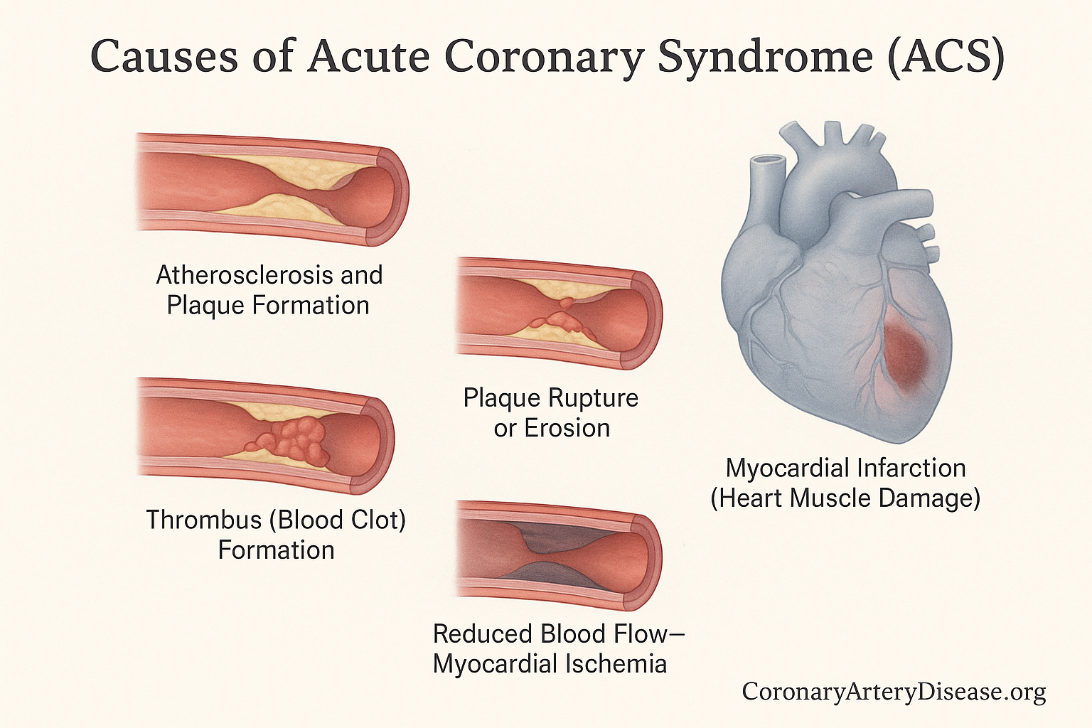
    First, here’s a quick list of the main mechanisms involved in the pathophysiology of Acute Coronary Syndrome

    • Atherosclerosis and Plaque formation
    • Plaque rupture or erosion
    • Thrombus (blood clot) formation
    • Reduced blood flow leading to myocardial ischemia
    • Heart muscle damage or death, resulting in myocardial infarction
    • Other causes, such as supply-demand mismatch (Type 2myocardial infarction), Spontaneous Coronary Artery Dissection (SCAD), or Myocardial Infarction with No Obstructive Coronary Artery Disease.

    The most common way Acute Coronary Syndrome develops is linked to atherosclerosis. This is a long-term process where the heart’s arteries, which are usually smooth and open, become stiff and narrow due to the build-up of fatty deposits, cholesterol, and other substances forming what’s called plaque. When this plaque becomes unstable, it can either rupture (break open) or erode (wear away). When this happens, the body’s natural response is to try and “fix” the injury by forming a thrombus, which is a blood clot, over the damaged area.

    This blood clot can suddenly block the artery, significantly reducing or completely stopping the blood flow to a part of the heart muscle. This sudden lack of oxygen and nutrients is what causes myocardial ischemia, leading to symptoms like chest pain. If the blockage isn’t quickly resolved, the heart muscle cells deprived of oxygen begin to die, leading to a myocardial infarction, or heart attack. This process is known as Type 1 myocardial infarction, which is usually what people refer to when they talk about a “heart attack”.


    However, not all heart attacks are caused by a sudden clot from plaque rupture or erosion. Sometimes, a heart attack, classified as Type 2 myocardial infarction, occurs due to a severe imbalance between the heart’s oxygen supply and its demand, without a direct sudden plaque-related blockage. This can happen if the heart needs a lot more oxygen (e.g., during extreme stress or a very fast heart rate) or if the body’s oxygen supply is critically low (e.g., from severe anemia or very low blood pressure). Other less common causes of Acute Coronary Syndrome include Spontaneous Coronary Artery Dissection, which is when a tear occurs in the wall of a coronary artery, creating a false channel that squeezes the main blood vessel and reduces blood flow. Another scenario is Myocardial Infarction with No Obstructive Coronary Artery Disease, where a heart attack is diagnosed, but angiography (a special X-ray of the heart’s arteries) doesn’t show significant blockages.

    Furthermore, recent insights indicate that infections like COVID-19 can also contribute to Acute Coronary Syndrome by causing direct or indirect inflammation and injury to the heart muscle, or by increasing the risk of blood clots. Understanding these different mechanisms is vital because treatment strategies may vary depending on the underlying cause.


    Other Similar Questions


    Resources

    • Bergmark BA, Mathenge N, Merlini PA, Lawrence-Wright MB, Giugliano RP. Acute coronary syndromes. Lancet. 2022 Apr 2;399(10332):1347-1358. doi: 10.1016/S0140-6736(21)02391-6. PMID: 35367005; PMCID: PMC8970581.
    • Smith JN, Negrelli JM, Manek MB, Hawes EM, Viera AJ. Diagnosis and management of acute coronary syndrome: an evidence-based update. J Am Board Fam Med. 2015 Mar-Apr;28(2):283-93. doi: 10.3122/jabfm.2015.02.140189. PMID: 25748771.
  • How do atherosclerotic plaques form in the heart arteries?

    Overview

    For a long time, we thought of what causes Coronary Artery Disease (CAD), which leads to heart artery blockages, mainly as a problem of too much cholesterol simply building up. However, How do atherosclerotic plaques form in the heart arteries in the last decade, has dramatically changed: we now view it fundamentally as an inflammatory disorder. This means that the formation of these blockages, called atherosclerotic plaques, involves a complex interaction between risk factors (like high cholesterol or high blood pressure), cells within your artery walls, and even blood cells. Crucially, inflammation plays a major role at every step.

    A key recent insight is the concept of “arterial remodeling.” This means that in many cases, plaques grow outwards first, expanding the artery wall rather than immediately narrowing the inside passage13. This “hidden” growth can make significant blockages hard to detect early on, as they might not cause symptoms until they become unstable or much larger


    In Details

    The process of atherosclerotic plaque formation, known as atherogenesis, is a detailed journey involving various steps and components:

    Initial Triggers and Endothelial Activation: It begins when the inner lining of your arteries, called the endothelium, encounters various irritants or risk factors. These can include substances from certain bacteria, high levels of fats (dyslipidaemia), hormones associated with high blood pressure (hypertension), products linked to high blood sugar (hyperglycaemia), or inflammatory signals from excess body fat. When the endothelium is exposed to these factors, its cells start to display “adhesion molecules” on their surface. These molecules act like sticky flags, encouraging certain white blood cells from your bloodstream—primarily immune cells called mononuclear phagocytes and T lymphocytes—to stick to the inner surface of the artery wall.

    Leukocyte Migration and Communication: Once these white blood cells adhere, they receive signals that help them move from the bloodstream into the inner layer of the artery, known as the intima. Inside the intima, these newly arrived immune cells begin to communicate with the artery’s own cells, including the endothelial cells and smooth muscle cells (SMCs). This communication involves a complex exchange of chemical messengers, such as various cytokines (proteins that mediate inflammation and immune responses), lipid mediators, and other substances that influence the artery’s behaviour. This interaction creates an “inflammatory ferment” within the early plaque.

    Smooth Muscle Cell Migration and Matrix Formation: A major consequence of this ongoing inflammation is the migration of smooth muscle cells (SMCs) from a deeper layer of the artery wall (the tunica media) into the intima. Once in the intima, these SMCs multiply and produce a rich and complex extracellular matrix, which is a kind of scaffolding material.

    Lipoprotein Trapping and Modification: Certain components of this matrix, particularly proteoglycans, can bind to lipoproteins (the carriers of cholesterol in your blood), prolonging their stay within the artery wall. This extended residence makes these lipoproteins more vulnerable to damage, such as oxidative modification or glycation (a non-enzymatic conjugation with sugars). These modified lipoproteins then sustain and propagate the inflammatory response within the developing plaque.

    Necrotic Core Formation and Plaque Progression: As the lesion progresses, cells can die, including lipid-laden macrophages, which are immune cells that have taken up a lot of fat. The death of these cells leads to the extracellular deposition of their contents, including substances that can trigger blood clotting, like tissue factor. This accumulation of extracellular lipid forms the classic, fatty “necrotic” core within the atherosclerotic plaque. Additionally, calcification, similar to bone formation, can occur within the plaque


    What is “arterial remodelling”?

    Arterial remodelling is the process where atherosclerotic plaques initially grow outwards, expanding the artery wall, rather than immediately growing inwards and narrowing the blood vessel. This means a significant amount of plaque can accumulate without causing a noticeable blockage that would be detected by angiography.

    Can plaques go away?

    While it’s not a complete “disappearance” in the sense of the artery becoming perfectly normal, aggressive management of risk factors can lead to the regression or shrinkage of atherosclerotic lesions. However, this shrinkage might occur internally within the artery wall, meaning the degree of narrowing seen on an angiogram might not significantly change, even as the plaque becomes less risky

    Is CAD just about blocked arteries?

    No, CAD is far more than just blocked arteries. It’s a complex, widespread inflammatory disease affecting the entire arterial system. While significant blockages can cause symptoms and require treatment, the underlying inflammatory process and the presence of numerous “hidden,” non-obstructive plaques are crucial to understanding and managing the disease


    Resources

    For more detailed information, you can refer to the source document:

    • Libby, P., & Theroux, P. (2005). Pathophysiology of Coronary Artery Disease. Circulation, 111(25), 3481–3488.