For individuals, whether you’re a patient, know someone with Coronary Artery Disease (CAD), or are simply interested, understanding The Diagnosis of Coronary Artery Disease is key.
Overview
Diagnosing Coronary Artery Disease typically begins with a healthcare professional assessing your symptoms, especially chest pain, and your individual risk factors. This initial assessment helps determine the likelihood that you have CAD. Based on this likelihood, various non-invasive tests, such as blood tests, electrocardiograms, and imaging scans, are used to gather more information about your heart’s health and blood flow. Invasive procedures, like coronary angiography, are generally reserved for situations where a treatment like revascularization is likely to be needed.
In Details
Diagnosing Coronary Artery Disease
- Blood tests
- Electrocardiogram (ECG or EKG)
- Echocardiogram
- Exercise stress test
- Nuclear stress test
- Heart CT scan (including CT coronary angiogram)
- Cardiac catheterization and angiogram
When you first see a healthcare professional, they will ask about your medical history and any symptoms you are experiencing, such as chest pain or shortness of breath. This initial assessment helps them estimate your pre-test probability – essentially, how likely it is that you have CAD before any major tests are done. For example, a general practitioner might use a tool called the Marburg Heart Score to calculate this probability based on factors like your age, sex, whether you have known vascular disease, if your symptoms occur during exertion, if the pain cannot be reproduced by touch, and if you believe the pain is heart-related. Specialists, like cardiologists, might use more detailed tables to determine this likelihood. If the estimated probability is very low (less than 15%), other causes for your symptoms will be considered first, and specific tests for CAD might not be necessary. If it’s very high (over 85%), CAD is often presumed, and treatment planning begins. For probabilities in between (15% to 85%), non-invasive tests are typically used.
As part of a basic evaluation, you might have a 12-lead resting ECG (Electrocardiogram), which checks the electrical activity of your heart. While a normal ECG doesn’t rule out CAD, abnormal patterns can indicate previous heart attacks or other issues. A resting echocardiogram, which uses sound waves to show blood flow through the heart, can also be considered to assess heart function and identify problems like weak areas that might suggest CAD.
For further evaluation, especially if your likelihood of CAD is moderate, your healthcare professional might recommend various non-invasive imaging techniques. These include:
• Exercise stress test:
This test checks your heart while you walk on a treadmill or ride a stationary bike, as symptoms often appear during physical activity. If you cannot exercise, medication can be given to simulate the effect of exercise on the heart.
• Nuclear stress test:
This uses a small amount of radioactive material, called a tracer, injected into your bloodstream to show how blood moves to your heart at rest and during activity, helping to find areas of poor blood flow or heart damage.
• Heart CT scan:
Other imaging techniques include stress echocardiography, myocardial perfusion SPECT (Single-photon Emission Computed Tomography), stress perfusion MRI (Magnetic Resonance Imaging), and dobutamine stress MRI. Most of these non-invasive tests have a sensitivity and specificity of around 85% for detecting obstructive CAD when compared to invasive coronary angiography.
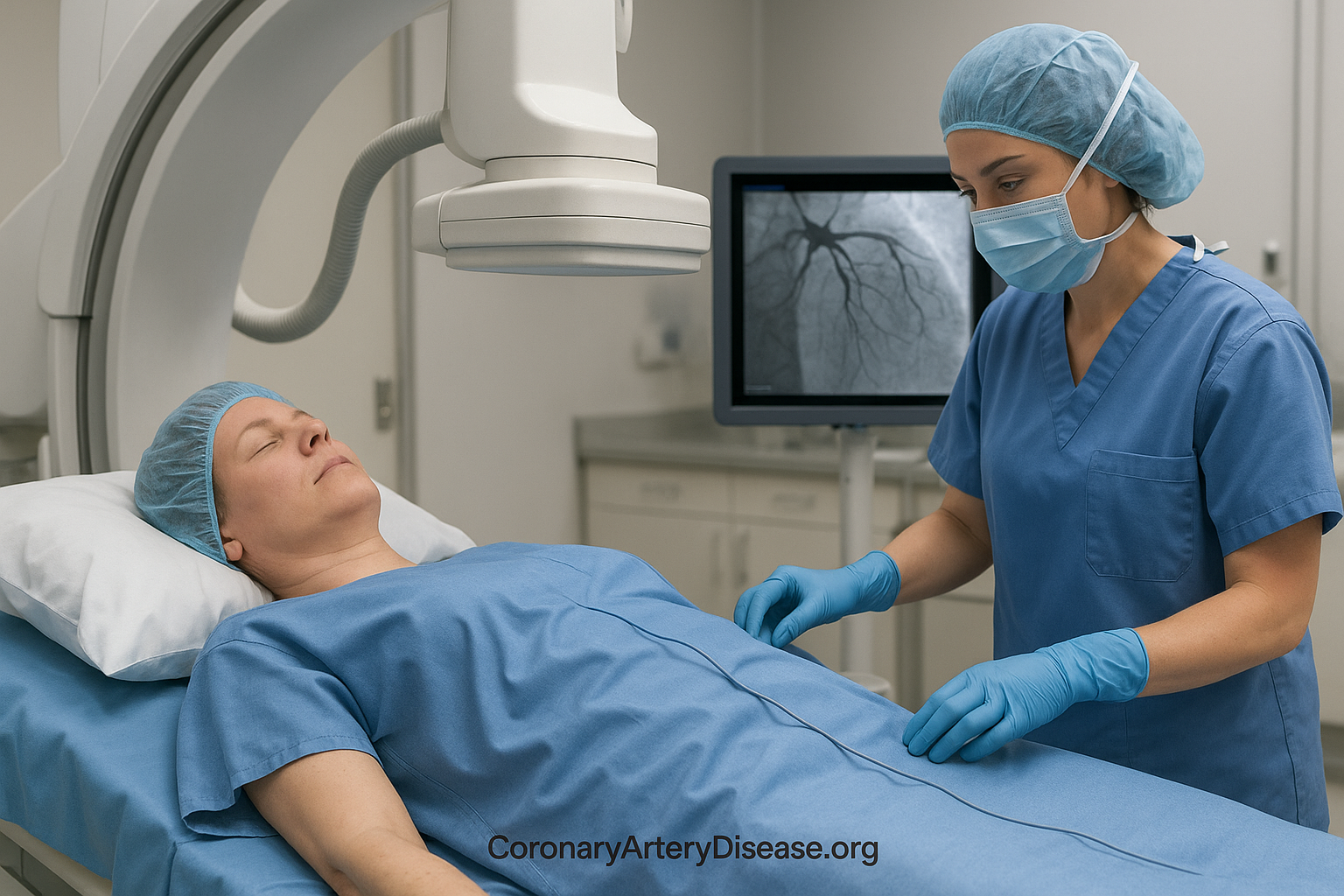
An invasive coronary angiography is a procedure where a long, thin tube (catheter) is inserted into a blood vessel and guided to your heart. A special dye is then injected to make your heart arteries visible on X-ray images, allowing doctors to see any blockages. This procedure is generally recommended only if the results are expected to lead to treatment, such as a revascularization procedure (like angioplasty or bypass surgery). It is not typically recommended if the probability of obstructive CAD is low, or if there are no signs of a problem after non-invasive tests
Resources:
For more detailed information, you can refer to the sources provided:
- Albus C, Barkhausen J, Fleck E, Haasenritter J, Lindner O, Silber S. The Diagnosis of Chronic Coronary Heart Disease. Dtsch Arztebl Int. 2017 Oct 20;114(42):712-719. doi: 10.3238/arztebl.2017.0712. PMID: 29122104; PMCID: PMC5686296.
- Coronary artery disease – Diagnosis and treatment – Mayo Clinic


Leave a Reply