Category: Uncategorized

  • Who is at high risk for Acute Coronary Syndrome?

    Who is at high risk for Acute Coronary Syndrome?

    Hello there, whether you’re a patient, someone who knows a patient, or just looking to understand more about heart health, let’s discuss who is at high risk for Acute Coronary Syndrome.


    Overview

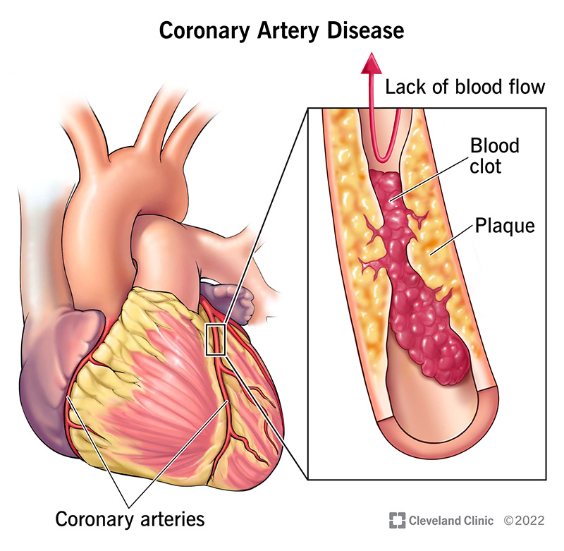
    When someone experiences an Acute Coronary Syndrome (ACS), like a heart attack, it’s typically caused by a blood clot forming in one of the heart’s arteries, blocking blood flow. This clot almost always happens because a fatty build-up in the artery wall, called an atherosclerotic plaque, becomes unstable and ruptures or erodes. Not all plaques are equally dangerous; some are particularly “rupture-prone” or “vulnerable.”

    Our understanding of what makes someone high-risk for ACS has evolved. While we used to focus mainly on the individual “culprit” plaque that caused the event, we now recognize that it’s often a more widespread problem within the arteries, involving many potentially vulnerable plaques and general inflammation throughout the body. We also understand that the “fluid phase” of a person’s blood – meaning factors circulating in the blood itself – can make them more prone to clotting, creating what’s known as a “vulnerable patient”. So, high risk isn’t just about one bad spot; it’s about the overall health of the arteries and the body’s clotting tendencies.


    In Details

    Let’s have a quick look at what characterize the vulnerable atherosclerotic plaques and patients

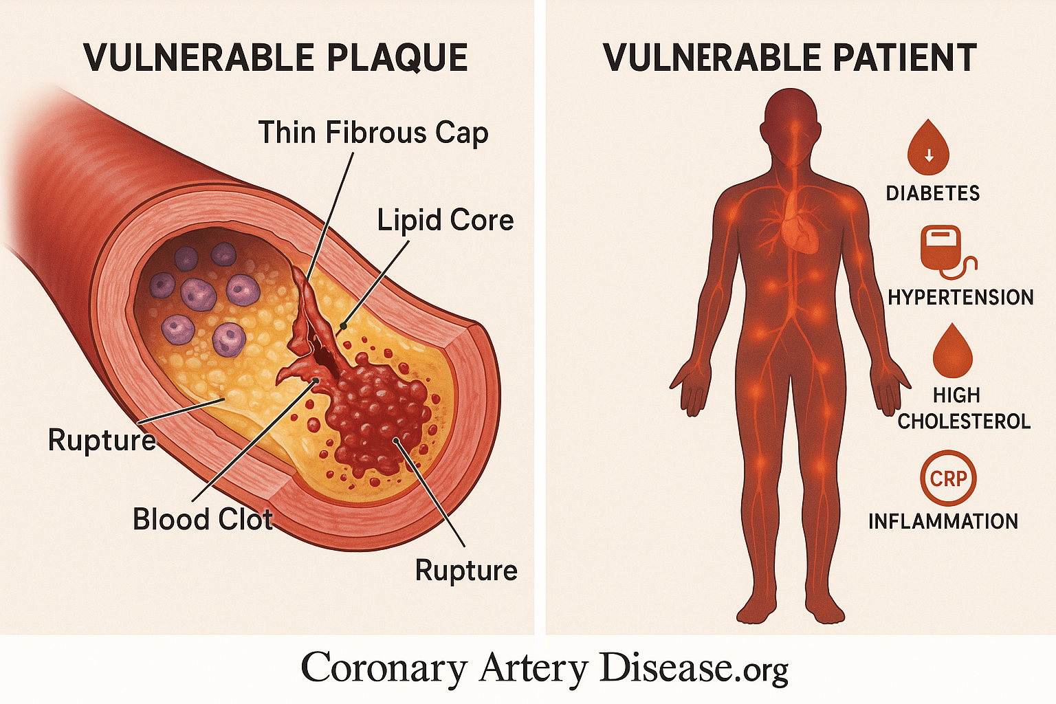
    • Presence of atherosclerotic plaques with a thin, fragile fibrous cap.
    • Plaques containing a large, soft lipid (fatty) core.
    • Plaques with a high number of inflammatory cells (e.g., macrophages).
    • Plaques with relatively fewer smooth muscle cells, which help strengthen the cap.
    • Widespread inflammation throughout the coronary arteries, not just at one site.
    • Circulating blood factors that promote clotting or hinder clot breakdown (e.g., high Plasminogen activator inhibitor-1 or PAI-1).
    • Presence of “hidden” plaques that have grown outward (compensatory enlargement) and don’t cause significant blockages but are still vulnerable.
    • Conditions like diabetes and obesity which can increase pro-clotting factors.

    A plaque that is “rupture-prone” or “vulnerable” possesses specific anatomical and cellular characteristics that make it susceptible to disruption. Primarily, these plaques are distinguished by a thin, fragile fibrous cap, which is the protective layer covering the fatty core. Beneath this cap lies a large, soft lipid core, rich in cholesterol and cellular debris. This core is particularly unstable. At a cellular level, these vulnerable plaques are heavily populated by inflammatory cells, such as macrophages, which contribute to weakening the fibrous cap by secreting enzymes that break down its structural components.

    Conversely, they tend to have fewer smooth muscle cells, which are crucial for maintaining the cap’s strength and integrity. The death of lipid-laden macrophages within the plaque can also lead to the release of tissue factor (TF), a powerful trigger for blood clotting, into the extracellular space. While fibrous cap rupture is the most common cause of acute coronary thrombosis, other mechanisms like superficial erosion of the artery lining, bleeding within the plaque (intraplaque hemorrhage), or erosion of a calcified nodule can also trigger a clot.

    The understanding of ACS has significantly shifted from viewing it as solely due to a single, critically narrowed artery or one “vulnerable plaque.” We now recognize that atherosclerosis is a widespread inflammatory disorder. Many plaques, even those that do not cause significant narrowing (known as “non stenotic lesions”), can be vulnerable. This is because arteries often undergo compensatory enlargement, meaning they grow outwards to accommodate the plaque without blocking blood flow, making the plaque “hidden” from detection by traditional angiography. Patients experiencing ACS often have multiple disrupted plaques throughout their coronary arteries, not just one “culprit lesion,” indicating a pan-coronary process driven by diffuse inflammation.

    This widespread inflammation in the arteries, alongside the specific characteristics of individual plaques, contributes to the overall risk. Furthermore, systemic factors in the “fluid phase” of the blood also play a critical role. For instance, high levels of Plasminogen activator inhibitor-1 (PAI-1) can reduce the body’s natural ability to dissolve blood clots, predisposing an individual to thrombosis. Conditions like diabetes and obesity can elevate PAI-1 levels, further contributing to a pro-clotting state. This collective understanding has led to the concept of the “vulnerable patient,” where overall systemic factors, combined with multiple vulnerable plaques, define the true risk of ACS.


    Other Similar Questions

    Can a person have many vulnerable plaques?

    Yes, studies show that patients with ACS often have multiple vulnerable plaques throughout their coronary arteries, not just one.

    What is the “no-reflow phenomenon”?

    This is when tiny pieces of a ruptured plaque or clot break off and travel downstream, blocking the very small blood vessels (microcirculation) in the heart muscle, even if the main artery has been opened.

    What is the dual-phase approach to treating acute coronary syndromes (ACS)?

    Beyond dealing with the immediate “culprit” lesion, the second phase focuses on “stabilizing” other plaques and reducing the patient’s overall vulnerability to future events. This means not just fixing the visible blockage, but also tackling the underlying, widespread issues like inflammation and the body’s tendency to form clots. This comprehensive strategy aims to protect against future acute events, which is crucial for long-term heart health.


    Resources

    For more detailed information, you can refer to the source document:

    • Libby, P., & Theroux, P. (2005). Pathophysiology of Coronary Artery Disease. Circulation, 111(25), 3481–3488.

  • How do atherosclerotic plaques form in the heart arteries?

    Overview

    For a long time, we thought of Coronary Artery Disease (CAD), which leads to heart artery blockages, mainly as a problem of too much cholesterol simply building up. However, in the last decade, our understanding has dramatically changed: we now view it fundamentally as an inflammatory disorder. This means that the formation of these blockages, called atherosclerotic plaques, involves a complex interaction between risk factors (like high cholesterol or high blood pressure), cells within your artery walls, and even blood cells. Crucially, inflammation plays a major role at every step.

    A key recent insight is the concept of “arterial remodelling.” This means that in many cases, plaques grow outwards first, expanding the artery wall rather than immediately narrowing the inside passage13. This “hidden” growth can make significant blockages hard to detect early on, as they might not cause symptoms until they become unstable or much larger


    In Details

    The process of atherosclerotic plaque formation, known as atherogenesis, is a detailed journey involving various steps and components:

    Initial Triggers and Endothelial Activation: It begins when the inner lining of your arteries, called the endothelium, encounters various irritants or risk factors. These can include substances from certain bacteria, high levels of fats (dyslipidaemia), hormones associated with high blood pressure (hypertension), products linked to high blood sugar (hyperglycaemia), or inflammatory signals from excess body fat. When the endothelium is exposed to these factors, its cells start to display “adhesion molecules” on their surface. These molecules act like sticky flags, encouraging certain white blood cells from your bloodstream—primarily immune cells called mononuclear phagocytes and T lymphocytes—to stick to the inner surface of the artery wall.

    Leukocyte Migration and Communication: Once these white blood cells adhere, they receive signals that help them move from the bloodstream into the inner layer of the artery, known as the intima. Inside the intima, these newly arrived immune cells begin to communicate with the artery’s own cells, including the endothelial cells and smooth muscle cells (SMCs). This communication involves a complex exchange of chemical messengers, such as various cytokines (proteins that mediate inflammation and immune responses), lipid mediators, and other substances that influence the artery’s behaviour. This interaction creates an “inflammatory ferment” within the early plaque.

    Smooth Muscle Cell Migration and Matrix Formation: A major consequence of this ongoing inflammation is the migration of smooth muscle cells (SMCs) from a deeper layer of the artery wall (the tunica media) into the intima. Once in the intima, these SMCs multiply and produce a rich and complex extracellular matrix, which is a kind of scaffolding material.

    Lipoprotein Trapping and Modification: Certain components of this matrix, particularly proteoglycans, can bind to lipoproteins (the carriers of cholesterol in your blood), prolonging their stay within the artery wall. This extended residence makes these lipoproteins more vulnerable to damage, such as oxidative modification or glycation (a non-enzymatic conjugation with sugars). These modified lipoproteins then sustain and propagate the inflammatory response within the developing plaque.

    Necrotic Core Formation and Plaque Progression: As the lesion progresses, cells can die, including lipid-laden macrophages, which are immune cells that have taken up a lot of fat. The death of these cells leads to the extracellular deposition of their contents, including substances that can trigger blood clotting, like tissue factor. This accumulation of extracellular lipid forms the classic, fatty “necrotic” core within the atherosclerotic plaque. Additionally, calcification, similar to bone formation, can occur within the plaque


    What is “arterial remodelling”?

    Arterial remodelling is the process where atherosclerotic plaques initially grow outwards, expanding the artery wall, rather than immediately growing inwards and narrowing the blood vessel. This means a significant amount of plaque can accumulate without causing a noticeable blockage that would be detected by angiography.

    Can plaques go away?

    While it’s not a complete “disappearance” in the sense of the artery becoming perfectly normal, aggressive management of risk factors can lead to the regression or shrinkage of atherosclerotic lesions. However, this shrinkage might occur internally within the artery wall, meaning the degree of narrowing seen on an angiogram might not significantly change, even as the plaque becomes less risky

    Is CAD just about blocked arteries?

    No, CAD is far more than just blocked arteries. It’s a complex, widespread inflammatory disease affecting the entire arterial system. While significant blockages can cause symptoms and require treatment, the underlying inflammatory process and the presence of numerous “hidden,” non-obstructive plaques are crucial to understanding and managing the disease


      Resources

      For more detailed information, you can refer to the source document:

      • Libby, P., & Theroux, P. (2005). Pathophysiology of Coronary Artery Disease. Circulation, 111(25), 3481–3488.

    • What causes Coronary Artery disease ?

      Overview

      For many years, doctors and scientists thought that Coronary Artery Disease (CAD), often called “heart artery disease,” was mainly caused by too much cholesterol building up in your blood vessels. While cholesterol certainly plays a role, our understanding has changed a lot. We now know that CAD is fundamentally an inflammatory condition, almost like an ongoing battle inside your arteries. This inflammation is crucial at every stage of the disease, from its very beginning to its progression, and even contributes to serious events like heart attacks

      .

      This new understanding means that managing CAD isn’t just about clearing blockages; it’s also about calming the widespread inflammation that affects your arteries. This inflammatory process can even make non-obstructive plaques, which might not cause symptoms, very dangerous

      In Details

      The inflammatory process in your arteries starts when the inner lining of these blood vessels, called the endothelium, encounters various “risk factors”. These can include high levels of unhealthy fats like LDL cholesterol, hormones linked to high blood pressure, substances associated with high blood sugar (like in diabetes), or even inflammatory signals from excess body fat. When the artery lining senses these stressors, it becomes “sticky,” expressing molecules that act like hooks. These hooks then grab white blood cells, such as immune cells called monocytes and T lymphocytes, which are circulating in your blood. Once attached, these white blood cells are drawn into the inner layer of the artery wall.

      Once inside the artery wall, these immune cells don’t just sit there; they become active participants in a complex inflammatory “conversation” with your artery’s own cells, like endothelial cells and smooth muscle cells. They exchange molecular messages, releasing various inflammatory mediators. These include small fatty molecules (like prostanoids and leukotrienes), other locally acting substances (like histamine), and particularly proteins called cytokines and complement components. These mediators further amplify the inflammatory response, turning it into a persistent state of irritation within the artery.

      A major consequence of this ongoing inflammation is the migration of smooth muscle cells (SMCs) from a deeper layer of the artery (the tunica media) into the inner lining. These SMCs then multiply and lay down a complex network of structural materials, forming what becomes part of the atherosclerotic plaque. In response to inflammatory signals, these cells also secrete enzymes called matrix metalloproteinases (MMPs), which can remodel or even break down parts of the artery’s structure. Components of this newly formed plaque can bind to lipoproteins, like cholesterol, making them more susceptible to damage, such as oxidation. These damaged lipoprotein products, in turn, continue to fuel and spread the inflammatory response, creating a self-perpetuating cycle of disease. As the plaque grows, dead, lipid-filled immune cells can accumulate, forming a soft, fatty core within the plaque.

      This inflammatory process isn’t just confined to one area; recent research shows that it’s often widespread throughout the arteries of individuals who experience acute coronary syndromes (like heart attacks). While some plaques grow inwards and create noticeable blockages, many others grow outwards, a process called “compensatory enlargement”. This outward growth means that a significant amount of disease can be present without causing narrowing that would be visible on standard angiography.

      These “hidden” lesions, particularly those with a thin outer fibrous cap and a large lipid core, are very prone to rupture. When such a plaque disrupts, it can trigger blood clot formation, leading to sudden events even if it hadn’t caused any symptoms before. Markers of inflammation, such as myeloperoxidase, have been found to be elevated even in areas of the heart not directly affected by a heart attack, indicating a widespread inflammatory state. This shifts our view from focusing solely on a single “vulnerable plaque” to considering the “vulnerable patient” with diffuse inflammation

      Other similar questions

      How do specific risk factors, like high blood pressure or diabetes, contribute to inflammation in arteries?

      When the inner lining of arteries, the endothelium, encounters risk factors such as high blood pressure (due to vasoconstrictor hormones) or high blood sugar (products of glycoxidation), these cells increase the expression of adhesion molecules

      What are the differences between a stable plaque and a “vulnerable” plaque, and how does inflammation play a role?

      Stable plaques, often those that cause significant narrowing (stenosis), typically have smaller lipid cores, more fibrous tissue, calcification, and thick fibrous caps2. In contrast, “vulnerable” plaques, which are prone to rupture and cause acute coronary syndromes (ACS), generally have large lipid cores, thin fibrous caps, and are populated by numerous inflammatory cells while lacking relatively in smooth muscle cells (SMCs)

      How do medications, such as statins, help by targeting inflammation, not just cholesterol levels?

      Statins and similar lipid-lowering therapies contribute to reducing recurrent coronary events by influencing the biology of the plaque, in addition to lowering cholesterol8. These successful therapeutic strategies appear to exert their benefit, at least in part, by combating inflammation8. Specifically, statins can reduce the blood levels of inflammatory markers like C-reactive protein

      Resources

      The information provided in this summary is based on the following scientific article:

      • Libby, P., & Theroux, P. (2005). Pathophysiology of Coronary Artery Disease. Circulation111(24), 3481–3488.

      This article provides a comprehensive review of the evolution in understanding the mechanisms of coronary artery disease. It is a valuable resource for deeper scientific understanding.

    • What is Coronary Artery Disease ?

      What is Coronary Artery Disease ?

      Overview

      Coronary Artery Disease (CAD) is a serious condition affecting the heart’s arteries. It’s often misunderstood, but our scientific understanding has greatly evolved. Previously, it was thought to be simply a build-up of cholesterol, like a plumbing problem where pipes get clogged. However, we now know that CAD is primarily an inflammatory disorder. This means that inflammation—the body’s natural response to injury or infection—plays a central role in every stage of the disease, from the very beginning of plaque formation to the sudden, serious events like heart attacks.

      CAD can show up in two main ways: as a long-term (chronic) condition, where symptoms might develop gradually, or as sudden, severe events (acute coronary syndromes like heart attacks or unstable angina). Importantly, these acute events often happen due to issues in blood vessels that weren’t even severely narrowed, challenging the old idea that only critically blocked arteries were dangerous. This new understanding means that treatment needs to go beyond just fixing blockages; it must also address the underlying inflammation and the overall health of your arteries.

      In Details

      At the heart of CAD is a process called atherogenesis, which is the formation of fatty plaques within the artery walls. This process starts when the inner lining of your arteries, called the endothelium, encounters various “risk factors”. These can include things like high cholesterol (dyslipidemia), high blood pressure, high blood sugar from diabetes, or even inflammatory signals from excess body fat. In response, the artery lining starts to express special “sticky” molecules that attract certain white blood cells, primarily immune cells called monocytes and T lymphocytes, from your blood. These cells then stick to the artery wall and move into the deeper layers. Once inside, these immune cells, along with the artery’s own cells, begin to communicate through inflammatory signals, setting the stage for plaque development.

      As this inflammatory process continues, muscle cells from the artery wall migrate into the inner lining and start to multiply. They also produce a complex mesh of proteins and other substances that form the main structure of the plaque. Certain parts of this mesh can trap cholesterol particles, making them more vulnerable to damage and further fueling the inflammatory response. Over time, calcium can also build up, making the plaque harder. As the plaque grows, fat-laden immune cells can die, releasing substances like tissue factor, which is crucial for blood clotting. All this accumulated fat and debris creates a soft, fatty core within the plaque, often called the “necrotic core”.

      One key insight is that for much of its life, an atherosclerotic plaque grows outwards, away from the centre of the artery. This is known as “compensatory enlargement”. This outward growth means that a significant amount of plaque can exist without narrowing the artery’s opening (the lumen). So, even if your angiogram (an X-ray of your blood vessels) looks relatively clear, you could still have widespread plaque build-up. These “hidden” lesions, which often have a thin protective outer layer (fibrous cap) and a large fatty core, might not cause any symptoms until they suddenly rupture and trigger a blood clot.

      Most acute coronary syndromes, like heart attacks, are caused by a sudden event: the disruption of one of these plaques, leading to the formation of a blood clot (thrombosis) that blocks blood flow. The most common way this happens is when the thin, protective fibrous cap over the plaque ruptures completely. Less commonly, the surface of the plaque might erode, or bleeding might occur within the plaque itself. When a plaque disrupts, it exposes materials like collagen and tissue factor (TF) inside the artery. These substances are powerful triggers for platelets (tiny blood cells involved in clotting) and the body’s clotting cascade, quickly forming a thrombus (blood clot) that can block the artery. Beyond the plaque itself, factors in your blood, such as substances that prevent clot breakdown (like PAI-1, which can be elevated in conditions like diabetes or obesity), can also increase your risk of dangerous blood clots.

      Comparisons

      Our understanding of CAD has shifted significantly:

      • From Cholesterol Storage to Inflammation: CAD was once primarily seen as a disease where cholesterol simply accumulated in artery walls. Now, it’s understood as an inflammatory disorder at its core, with inflammation driving plaque formation and progression.
      • From Narrowing to Widespread Disease: Doctors used to focus heavily on how much the arteries were narrowed (stenosis), which was easily seen on angiograms. However, we now know that such narrowings are often just the “tip of the iceberg”. Most plaques don’t cause significant narrowing due to “compensatory enlargement” (outward growth). This means CAD is often a widespread disease affecting many arteries, not just a few isolated spots.
      • From “Vulnerable Plaque” to “Vulnerable Patient”: Initially, there was a search for a single “vulnerable plaque” that was prone to rupture. While certain plaque characteristics (thin cap, large fatty core) do make them risky, we now recognise that patients prone to acute events often have multiple such plaques and widespread inflammation throughout their arterial system. This shift emphasizes that the “vulnerable patient” (someone with overall risk factors and diffuse arterial inflammation) is as important as, if not more important than, identifying just one problematic plaque.
      • From Symptom Relief to Plaque Stabilisation: Traditional treatment for chronic CAD focused on relieving symptoms by improving blood flow, often through procedures like angioplasty or bypass surgery. While these are still vital, the modern approach also prioritises “stabilising” other plaques and reducing overall cardiovascular risk, not just treating the most obvious blockages. This involves aggressive management of risk factors and systemic therapies that combat inflammation.

      Other Similar Questions

      • How is CAD managed or treated now? Current management involves not only procedures like angioplasty or bypass surgery to open severely blocked arteries but also crucial long-term strategies. These include aggressive management of modifiable risk factors (like blood pressure, cholesterol, and diabetes) through lifestyle changes and medications (such as statins, aspirin, and ACE inhibitors). The goal is to rapidly stabilise plaques and reduce overall inflammation to prevent future acute events.
      • Can CAD be prevented or reversed? Lifestyle measures, such as a healthy diet, regular exercise, and not smoking, are the foundation for preventing CAD. Aggressive management of risk factors has been shown to slow disease progression and can even lead to some regression of atherosclerosis. While plaques might not always shrink visibly on angiograms, studies suggest that they can become more stable and less prone to rupture, which is a key aspect of “reversibility”.
      • What are biomarkers, and how do they relate to CAD? Biomarkers are measurable substances in the blood that can indicate disease activity. In CAD, certain inflammatory markers, like C-reactive protein, can predict the risk of future heart events. Research is ongoing to see if monitoring these markers can help guide treatment, especially given that some medications, like statins, have anti-inflammatory effects independent of their cholesterol-lowering actions.

      Resources

      The information provided in this summary is based on the following scientific article:

      • Libby, P., & Theroux, P. (2005). Pathophysiology of Coronary Artery Disease. Circulation, 111(24), 3481–3488.

      This article provides a comprehensive review of the evolution in understanding the mechanisms of coronary artery disease. It is a valuable resource for deeper scientific understanding.